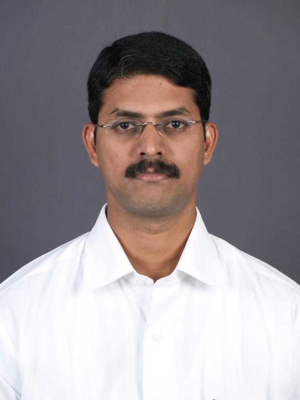
PROF. DR. K. MUGUNDHAN
Director & Professor
MD, DM (Neurology). Director and Professor of Neurology at the Institute of Neurology, Madras Medical College. He has been practicing in the field of Neurology since 2006 with a strong commitment to clinical care. His areas of interest include epilepsy, stroke, and demyelinating disorders. He is known for his excellent administrative skills. He is also deeply passionate about clinical teaching and mentoring young medical professionals.
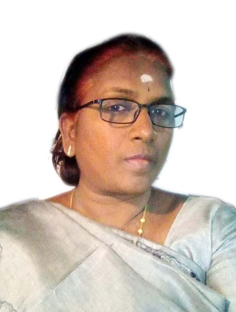
PROF. DR. N. THAMIL PAVAI
Professor
MD, DM (Neurology). Professor of Neurology at the Institute of Neurology, Madras Medical College. Member of IAN, IEA ,MDSI & IHS. Areas of interest movement disorders & cognitive neurology. ICMR Projects : 1) As a CO–PI in All India Multiple Sclerosis Registry. 2) As a CO–PI in Profile of Neuronal Damage based on clinical, immunological, imaging and spectroscopic alterations in patients recovered from SARS-CoV2 infection - A Cohort Study.
PROF. DR. R. VIVEKA SARAVANAN
Professor
MD, DM (Neurology). Professor of Neurology at Institute of Neurology, Madras Medical College. Experienced neurologist with a strong commitment to clinical care and academic excellence. Areas of interest include general neurology, neuroimmunology and epilepsy. He has a proven track record of guiding medical trainees and fostering professional development.
PROF. DR. N. SHANMUGA SUNDARAM
Professor
MD, DM (Neurology). Professor in Institute of Neurology, Madras Medical College. He is an experienced neurologist with areas of interest in general neurology, demyelinating disorders and electrophysiology. He has a strong commitment to comprehensive neurological care with emphasis on patient empathy.
DR. E. UMA MAHESHWARI
Associate Professor
MD, DM (Neurology). Assistant Professor in Institute of Neurology Madras Medical College. She has been practicing in the field of Neurology since 2013. Her areas of interest are demyelinating disorders and CNS Tuberculosis. She is also a co-principal investigator in ICMR Project - IN SHORT TB Meningitis trial and TBM Registry.
DR. L. A. RAVI
Associate Professor
MD, DM (Neurology). Assistant Professor in Institute of Neurology, Madras Medical College. He has been practicing in the field of Neurology since 2014 with a strong commitment to clinical care. His areas of interest include epilepsy, stroke, and demyelinating disorders. He is also deeply passionate about clinical teaching and mentoring young medical professionals.
DR. A. MARIAN JUDE VIJAY
Assistant Professor
MD, DM (Neurology). Assistant Professor in Institute of Neurology, Madras Medical College. He has been practicing in the field of Neurology since 2014. His areas of interest include epilepsy, stroke, demyelination and electrophysiology.
DR. V. NANDAKUMAR
Assistant Professor
MD, DM (Neurology). Assistant Professor at the Institute of Neurology, Madras Medical College. He has been practicing in the field of Neurology since 2016. He is CO–PI for ICMR Project – Hospital based stroke registry. His areas of interest include pediatric neurology and demyelinating disorders.
DR. E. NATARAJAN
Assistant Professor
MD, DM (Neurology). Assistant Professor at the prestigious Institute of Neurology, Madras Medical college. He has immense interest in clinical neurology. His other areas of interest include movement disorders, stroke care and neuromuscular disorders.
DR. V. RAJAGEMBEERAN
Assistant Professor
MD, DM (Neurology). Assistant Professor at the prestigious Institute of Neurology, Madras Medical college. He areas of interest include neurosonology, pediatric neurology, stroke and epilepsy.
DR. A. VIGNESH
Assistant Professor
MD, DM (Neurology). Assistant Professor at Institute of Neurology, Madras medical College. He is a dedicated neurophysician who has been practicing Neurology since 2017. He has also got his Speciality certification in Neurology in 2018. Since then, he is working as faculty of neurology and is passionate about teaching and developing clinical skills.
DR. V. PRAKASH
Assistant Professor
MD, DM (Neurology). Assistant professor at Institute of Neurology, Madras Medical College with expertise in stroke treatment and neurocritical care. He is practicing in the field of neurology since 2024. He is passionate about improving lives through comprehensive neurological care.
DR. SHYLAJA
Senior Resident
MD, DM (Neurology). Senior Resident at the Institute of Neurology, Madras Medical College. She is passionate about intellectual conglomeration, guiding and mentoring young prodigies. Her areas of interest include neurocritical care and neuroelectrophysiology.
DR. AUBIN MATHEW V
Senior Resident
DNB, DM (Neurology). Senior Resident at Institute of Neurology, Madras Medical College. His areas of interest include epilepsy, stroke, and neurocritical care.