1934: Founded as Goschen Institute of Pathology; inaugurated on March 26 by Sir George Fredrick Stanley.
Named after George Joachim Goschen, Governor of Madras (1924–1929) and Viceroy of India (1929–1931).
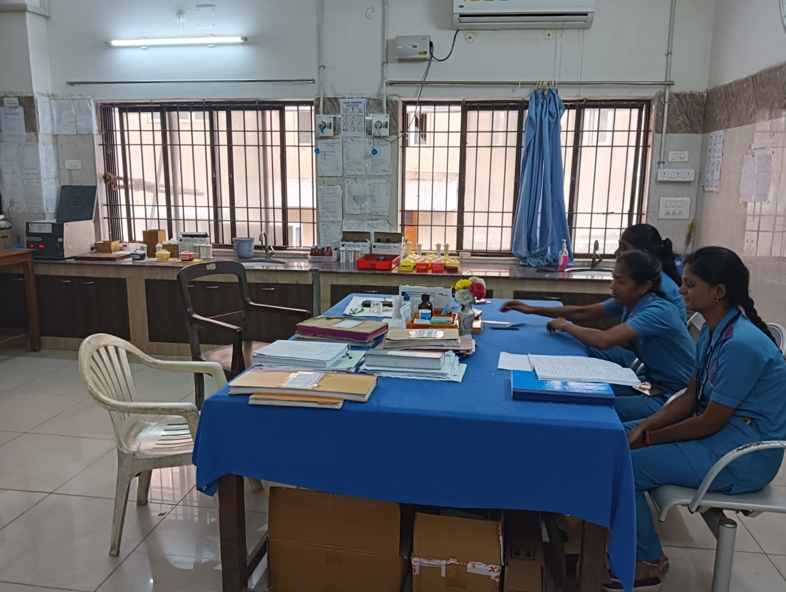
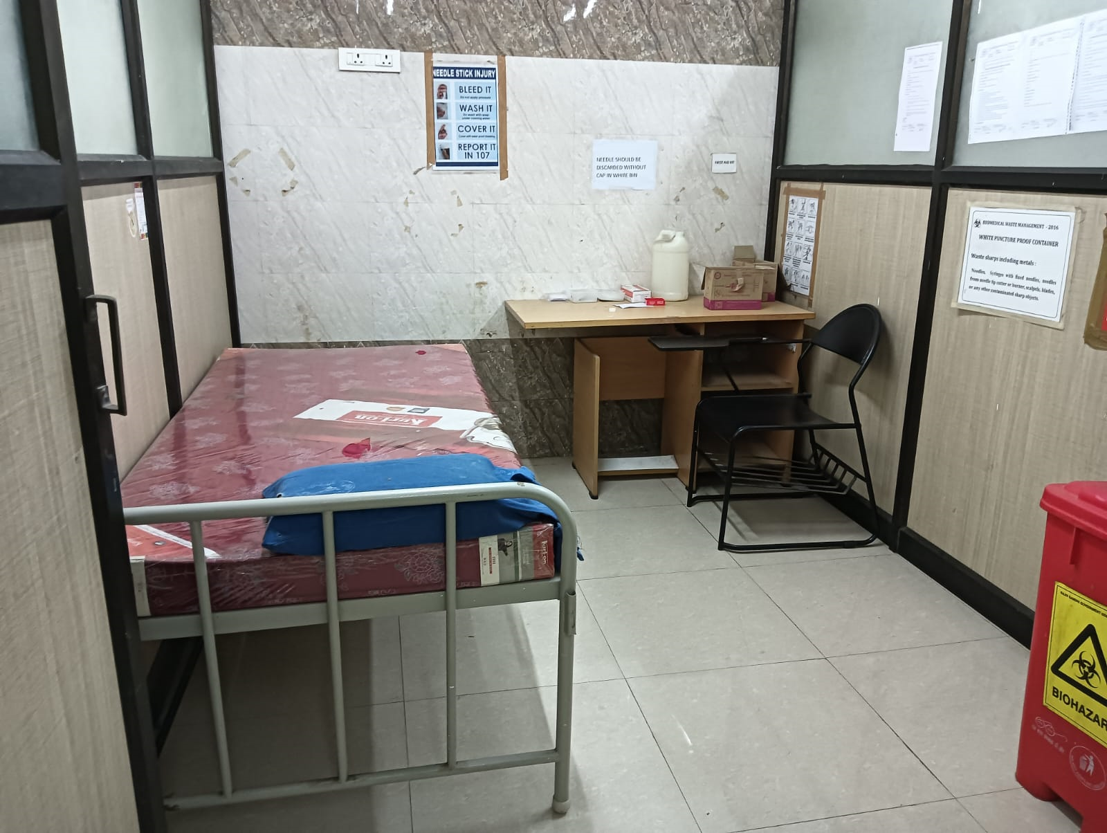
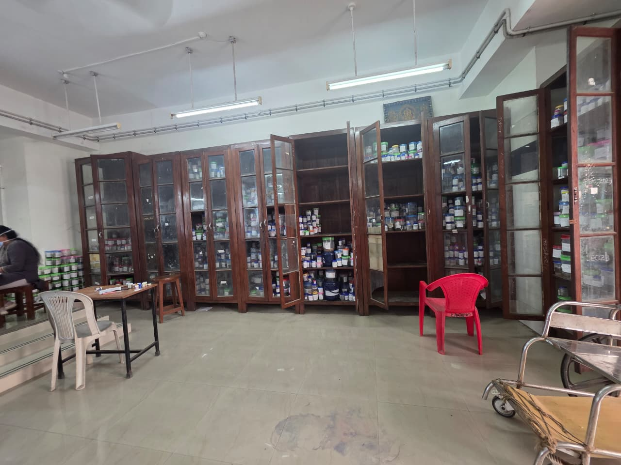
Initially located in what is now called the Goschen Block; currently in Tower Blocks 1 & 3,RGGGH and college
building 3rd floor.
Academic & Institutional Milestones:
1936: First chair in Pathology created; Prof. Srinivasulu Naidu became the first HOD.
1956: Exclusive MD Pathology program introduced under Medical Council of India.
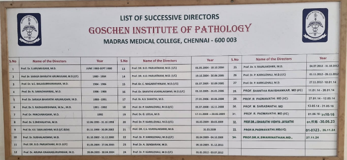
1980: Upgraded to Institute of Pathology; Prof. S. Arumugam appointed as first Director.
2010: PG seats increased from 4 to 10 MD; 8 Diploma seats added.
2019: MD seats increased from 10 to 18; DCP seats converted to MD.
MD Pathology students have been university toppers.
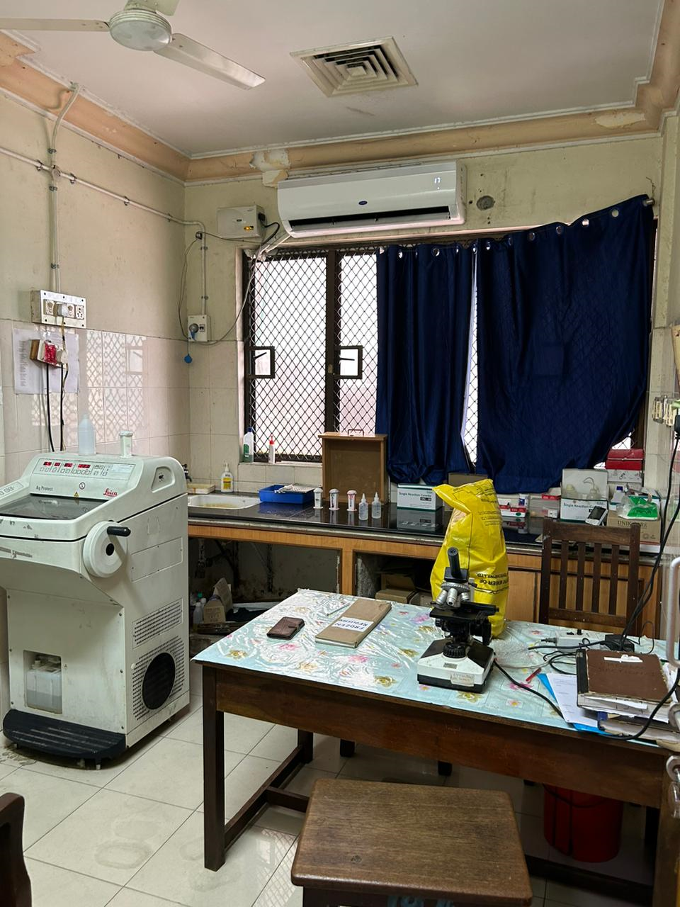
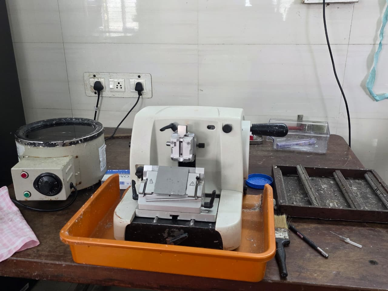
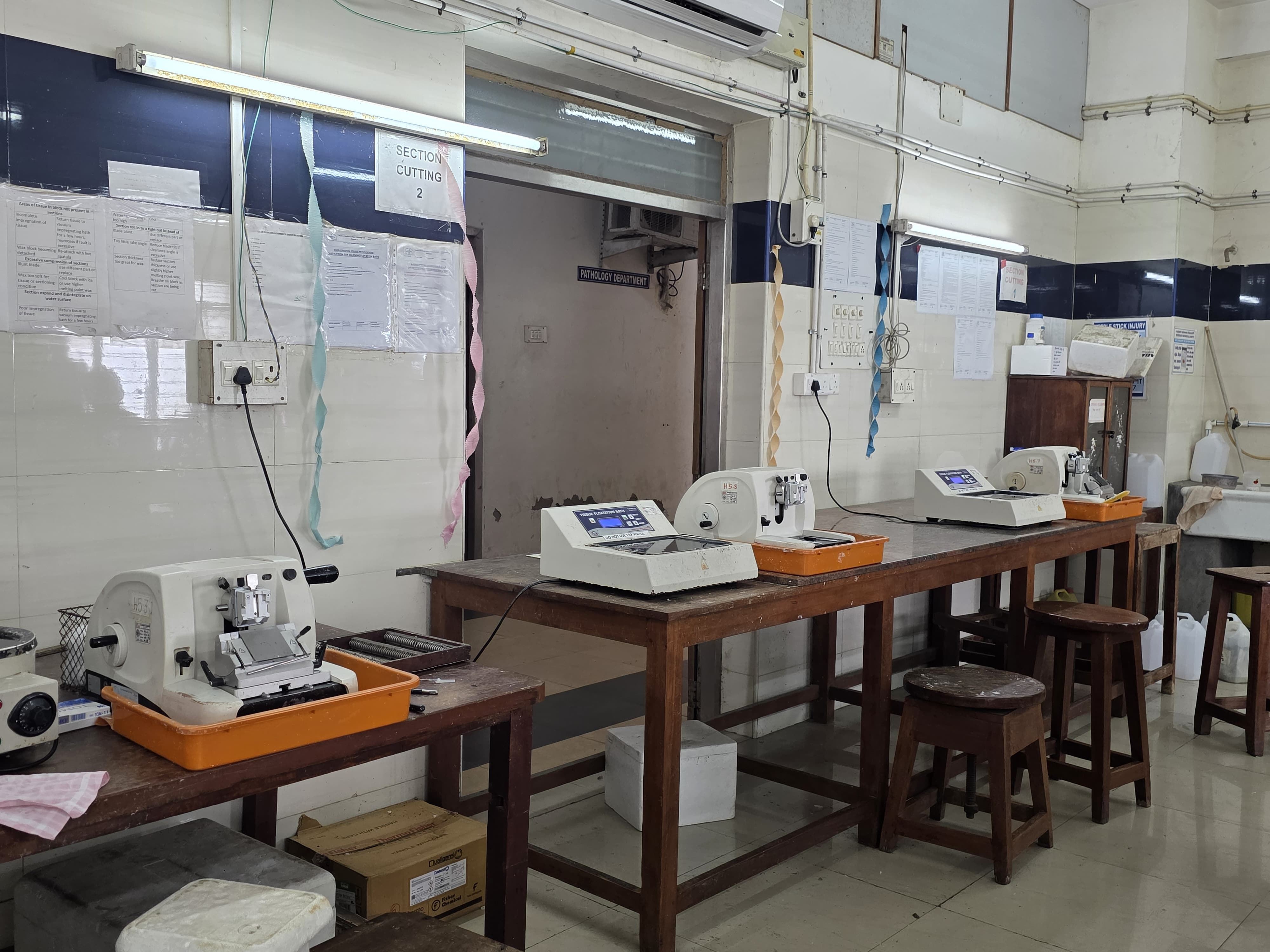
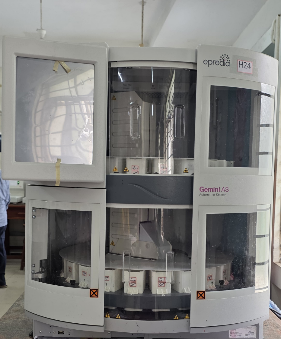
Diagnostic and Technological Advancements:
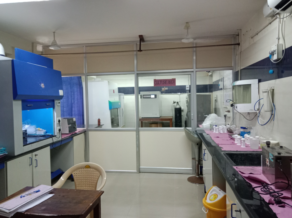
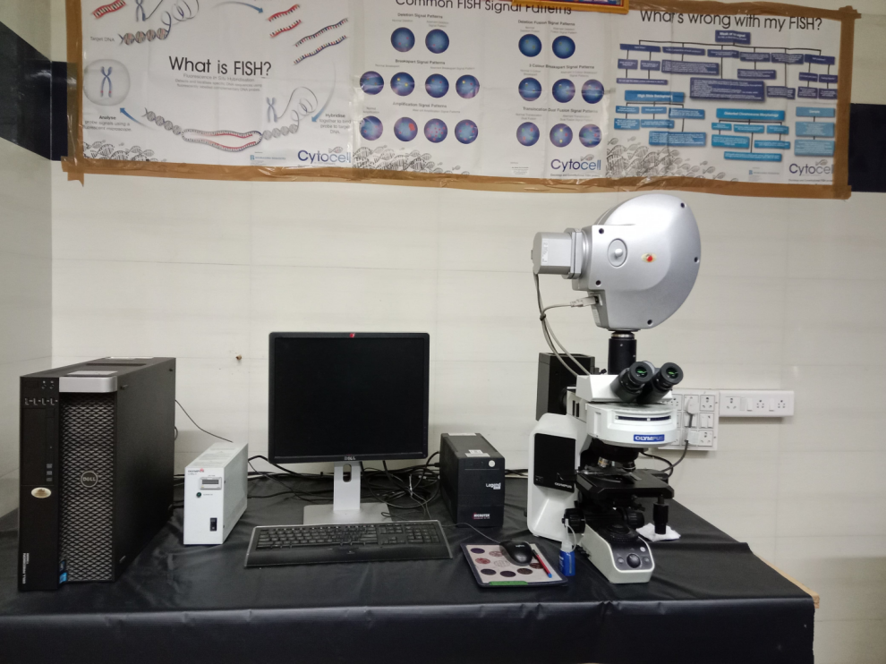
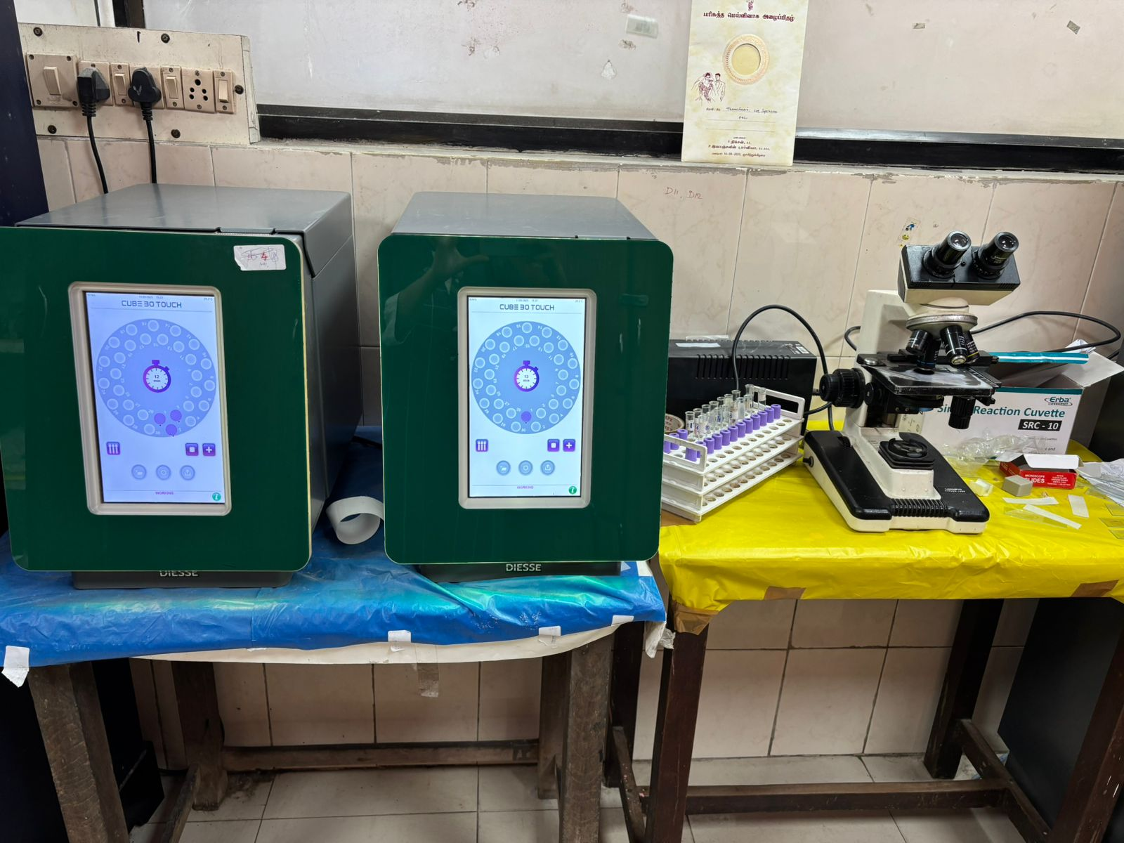
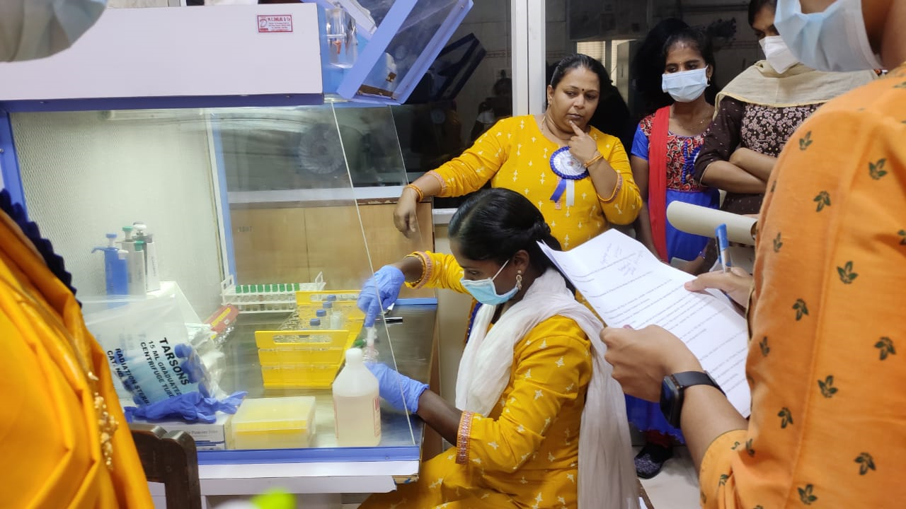
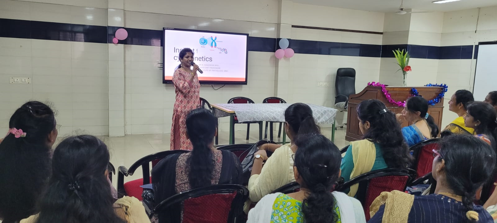
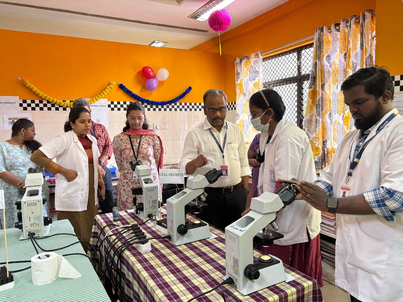
2015: Cytogenetics workstation commissioned
2016: PAP smear screening began under Master Health Checkup.
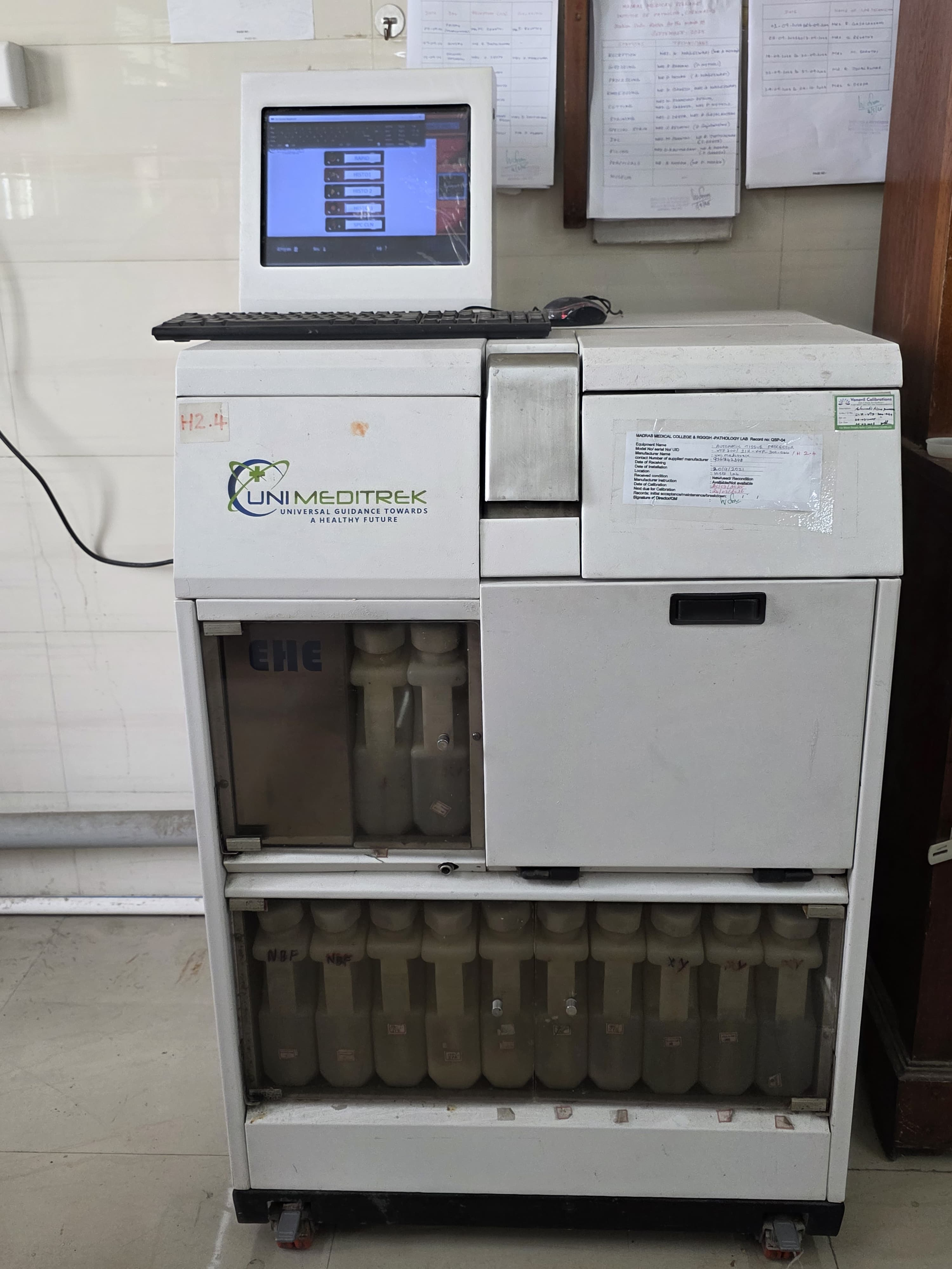
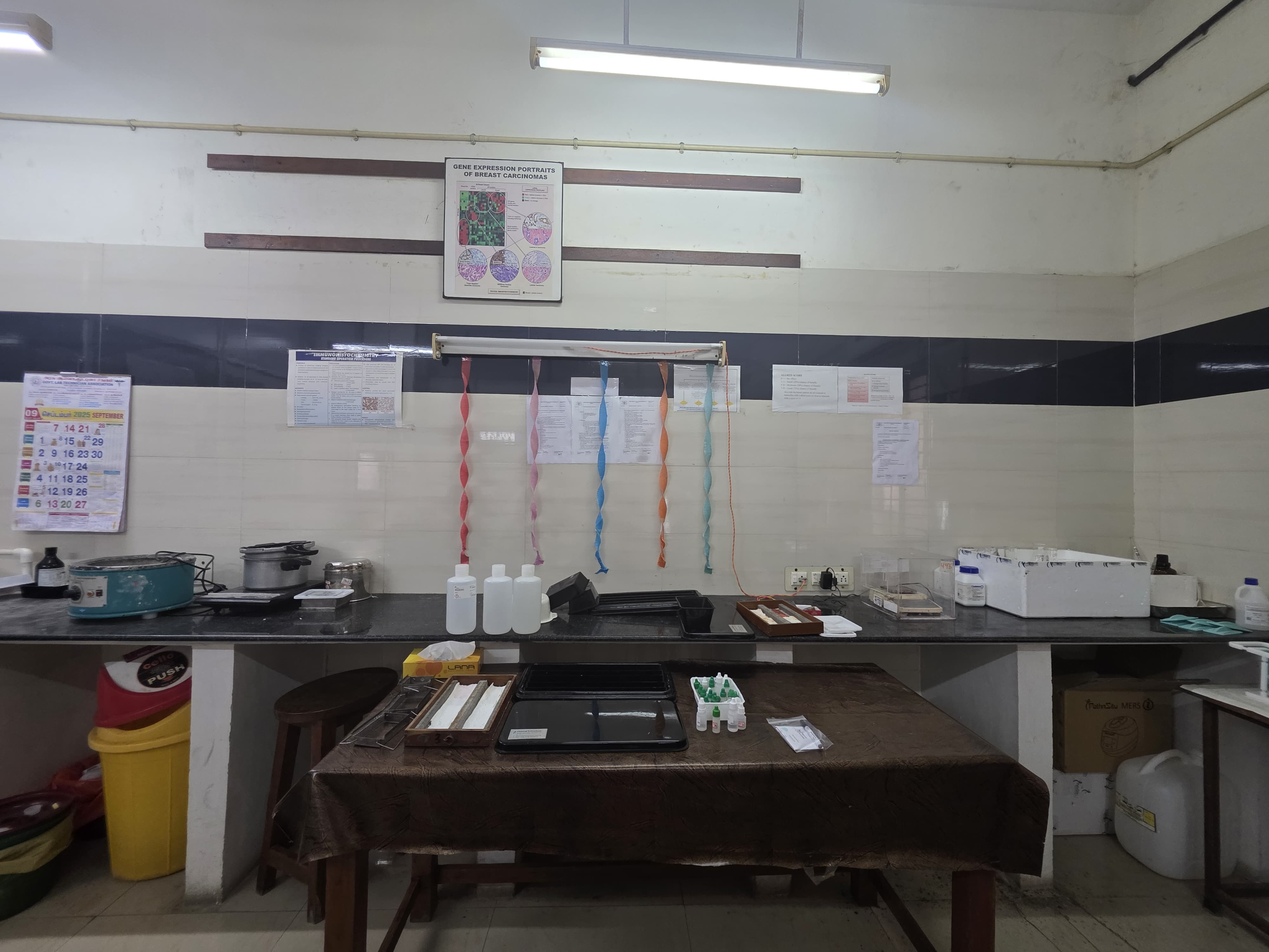
2017: Full implementation of Immunofluorescence and Immunohistochemistry (152 markers) including epithelial,
mesenchymal, lymphoma, and neuroendocrine panels.
2010: PG seats increased from 4 to 10 MD; 8 Diploma seats added.
2019: MD seats increased from 10 to 18; DCP seats converted to MD.
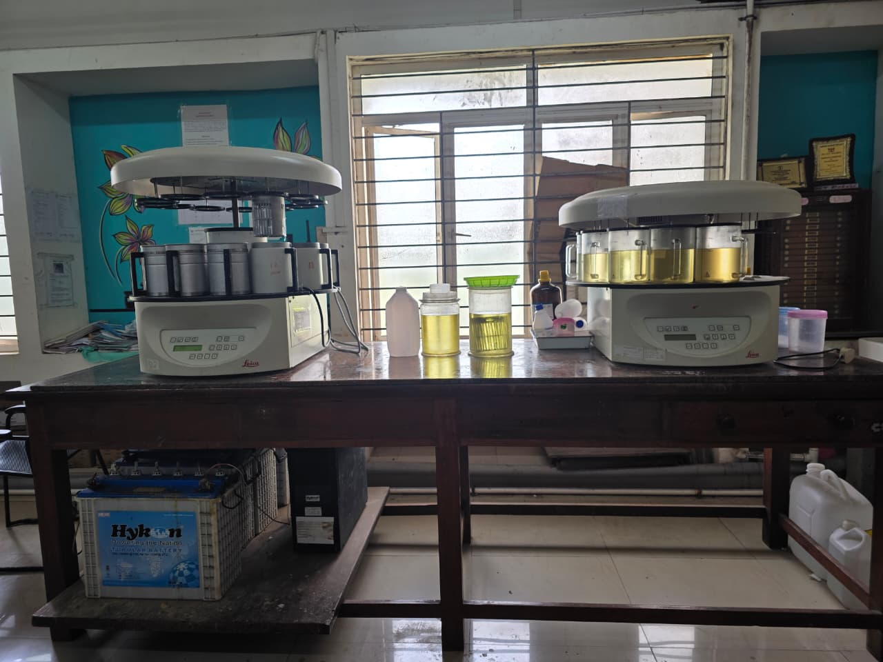
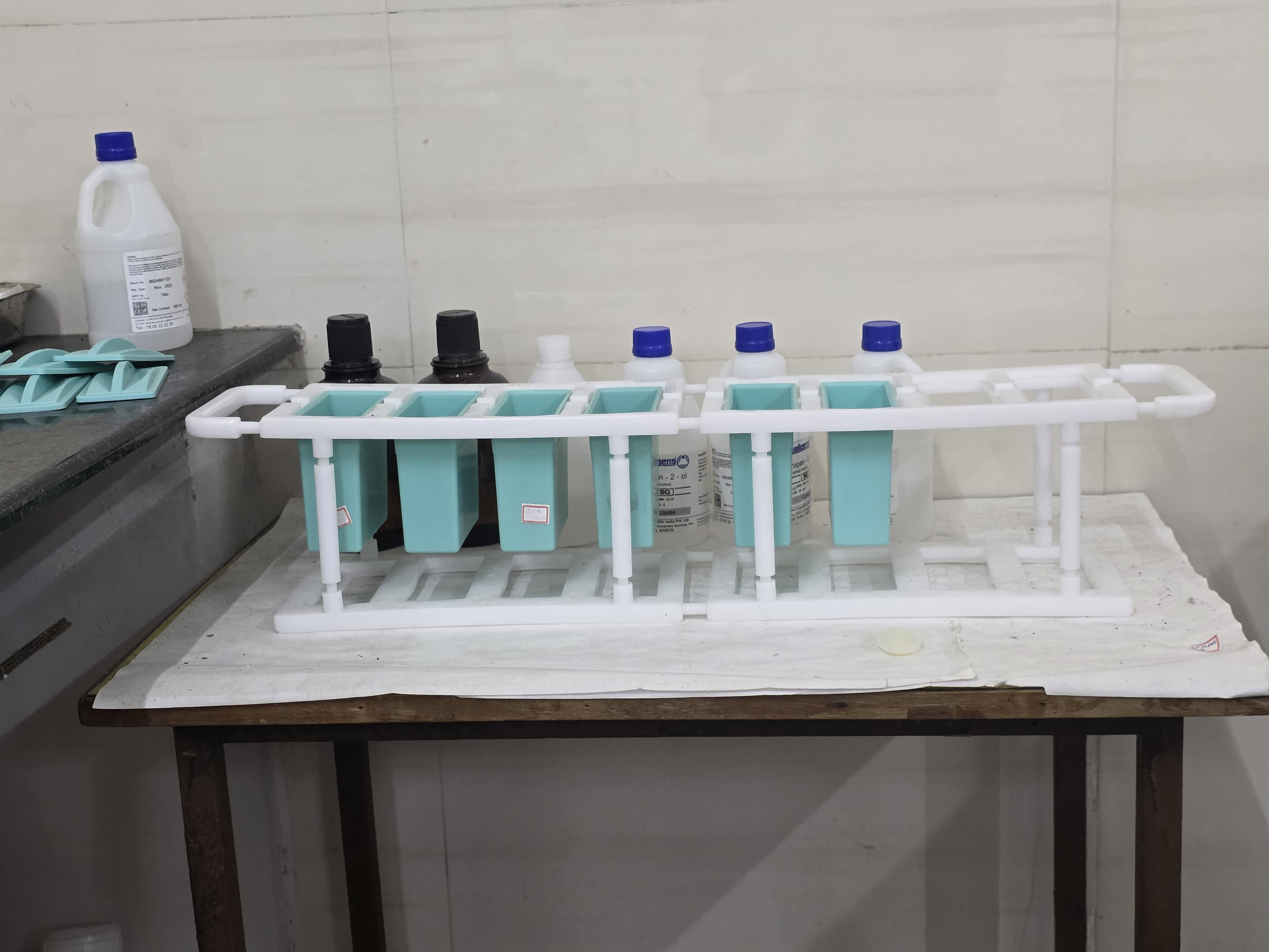
Bone marrow examination routinely done with special stains and cytogenetics as needed.
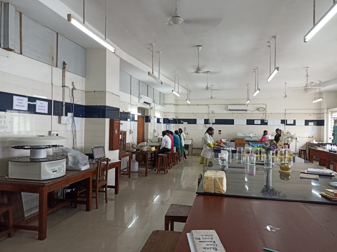
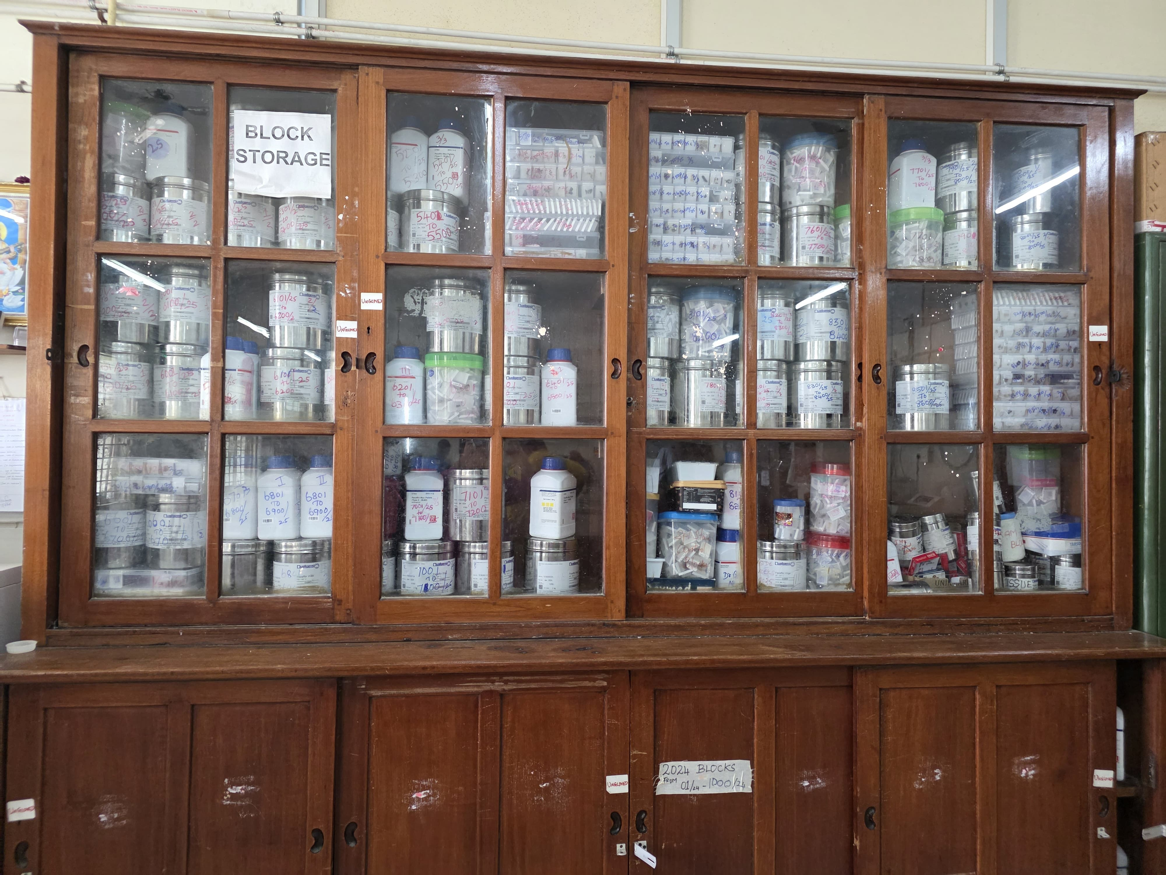
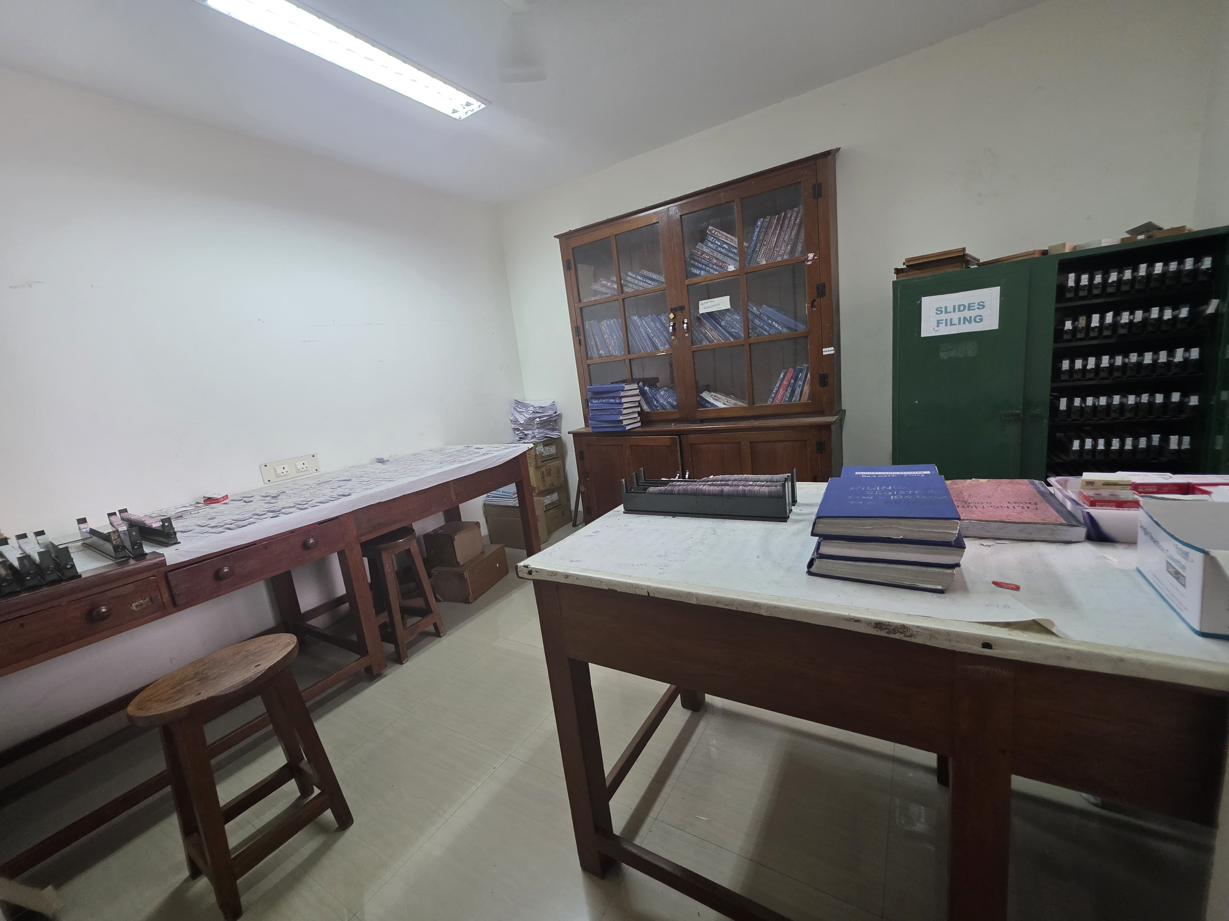
Museum houses 1568 mounted specimens, including rare one.
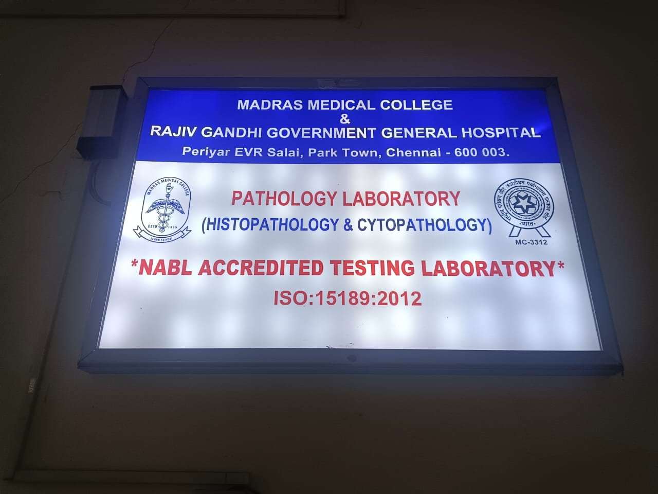
Recognition & Accreditation:
Feb 4, 2020: NABL accreditation (ISO 15189:2012) for Histopathology and Cytology , later extended to Hematology,
special stains and ER/PR/HER2 neu panel for breast
Feb 2024: NABL accreditation extended to all domains in pathology except cytogenetics.
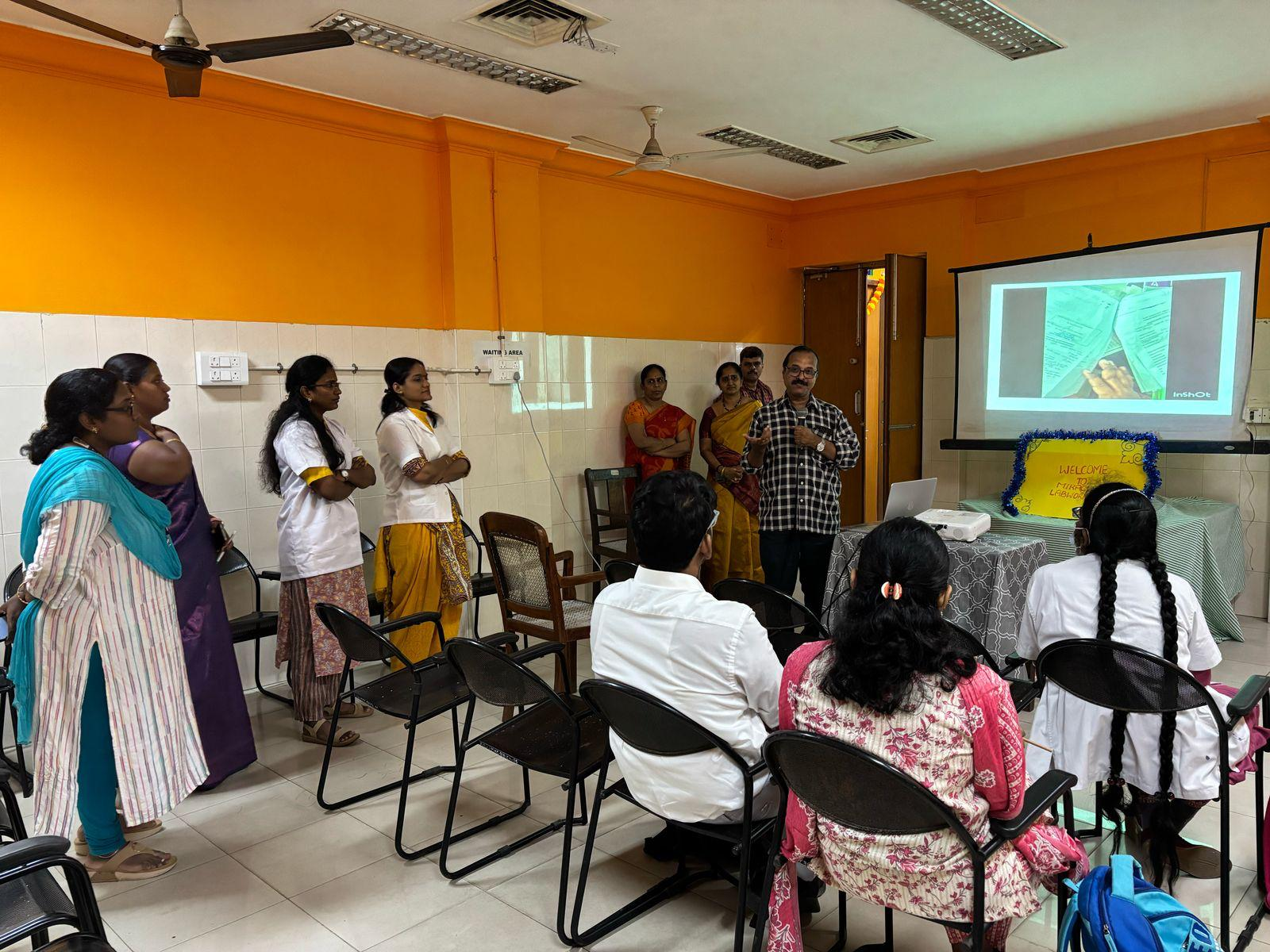
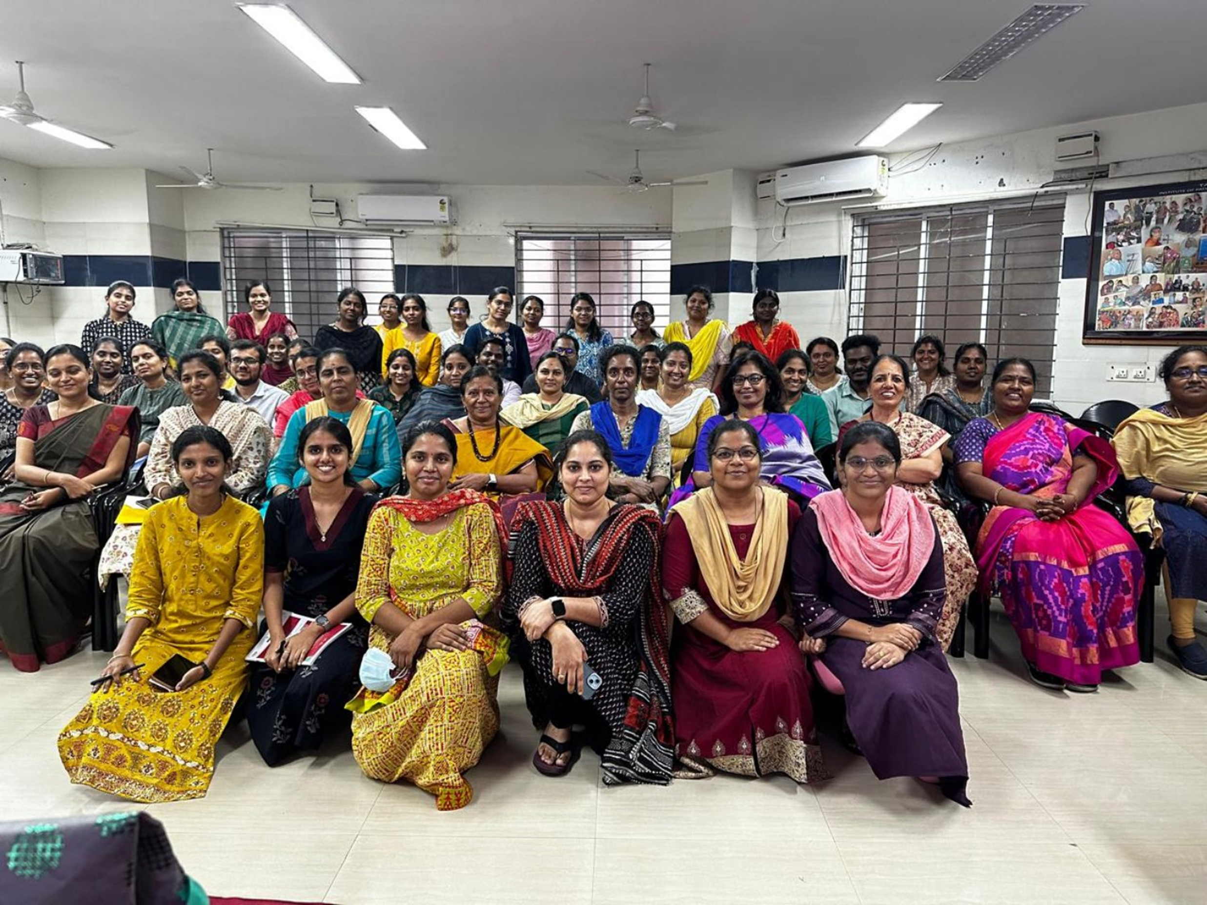
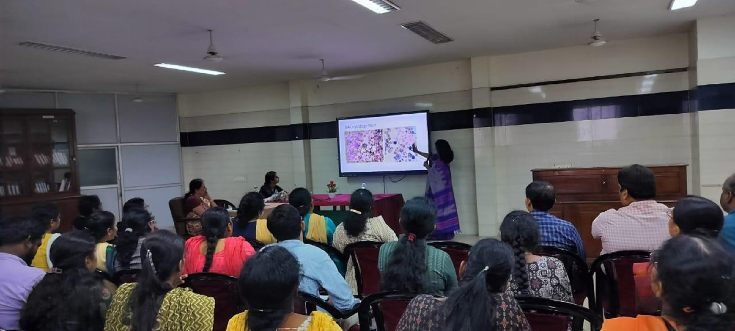
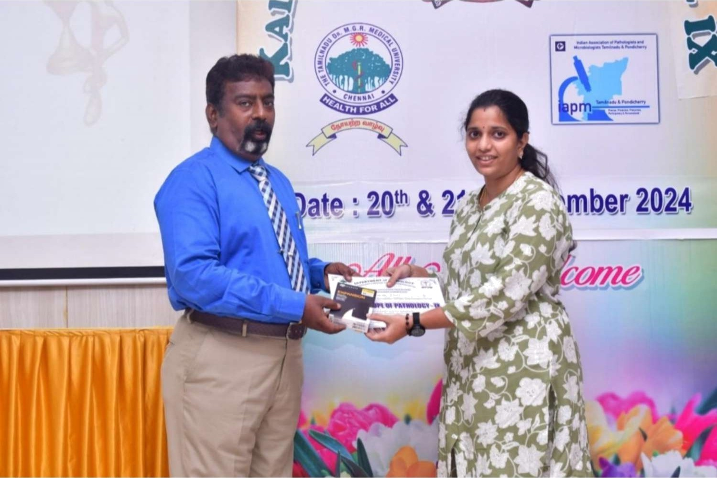
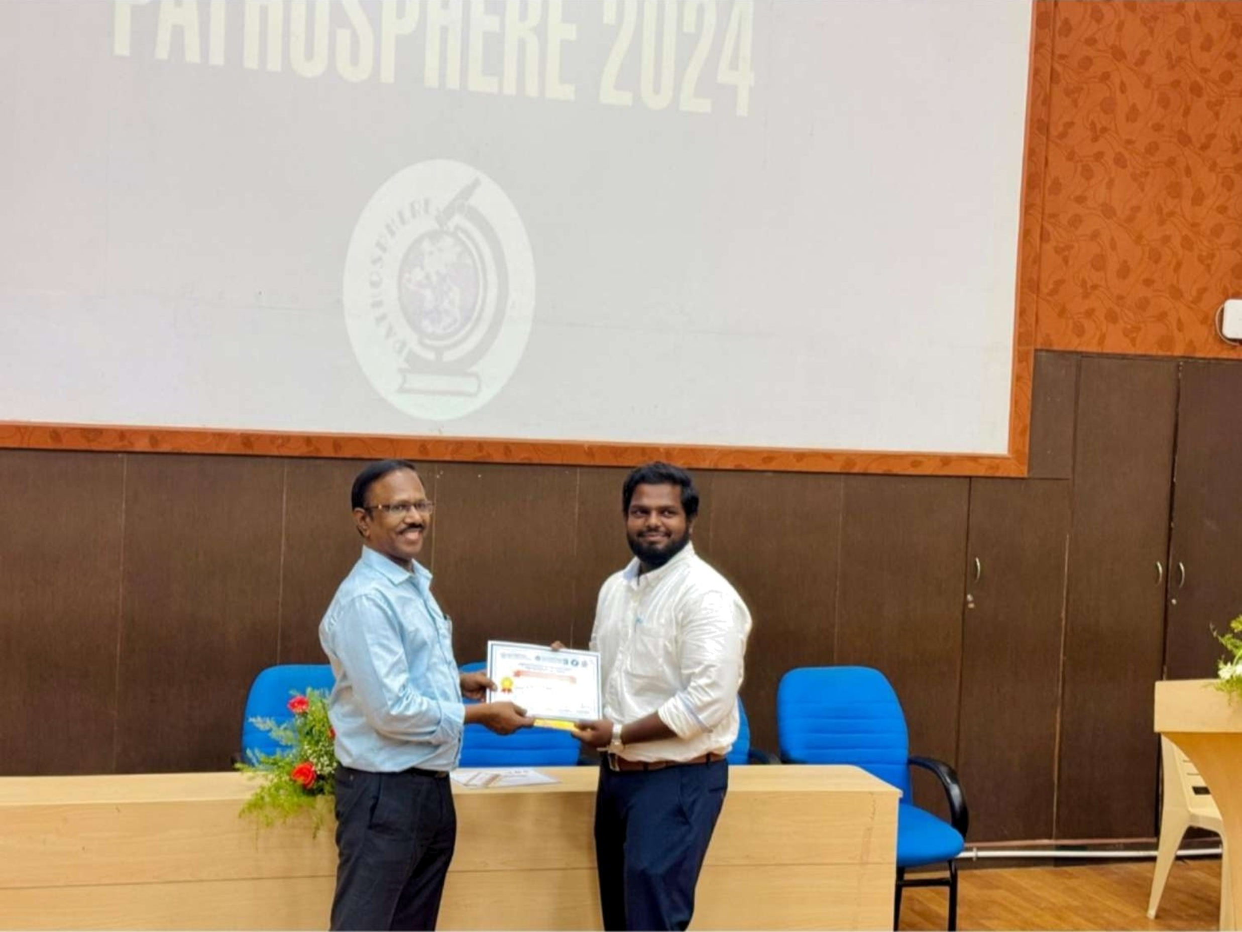
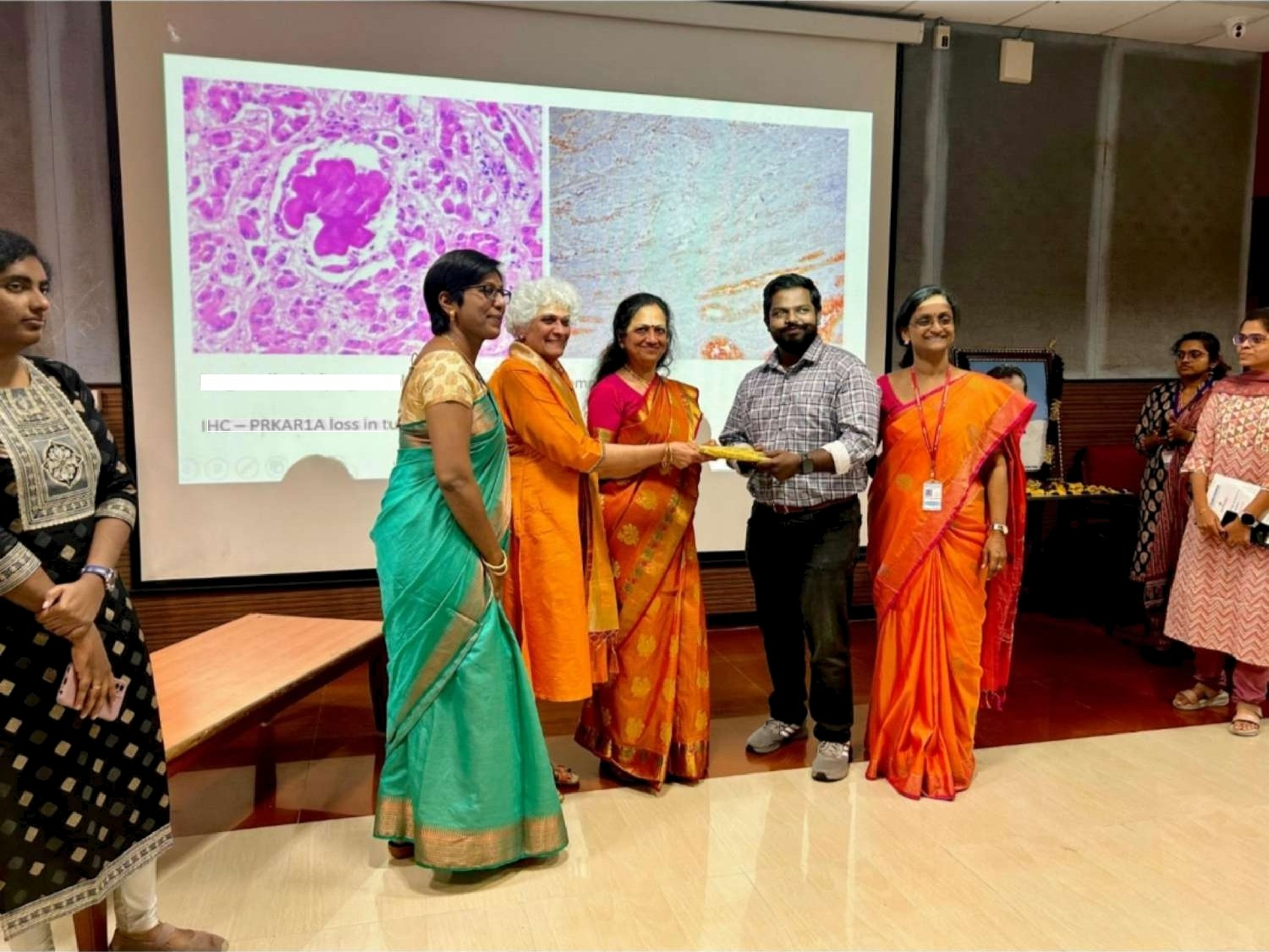
Workshops & CMEs:
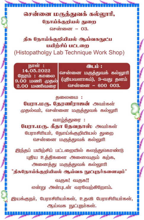
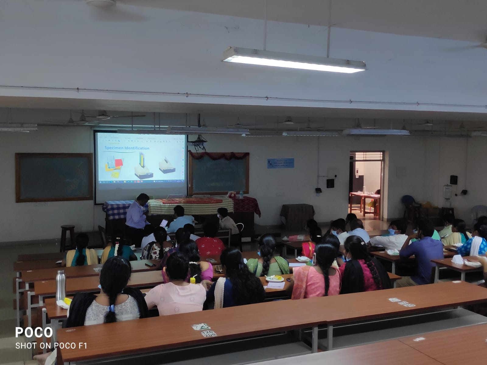
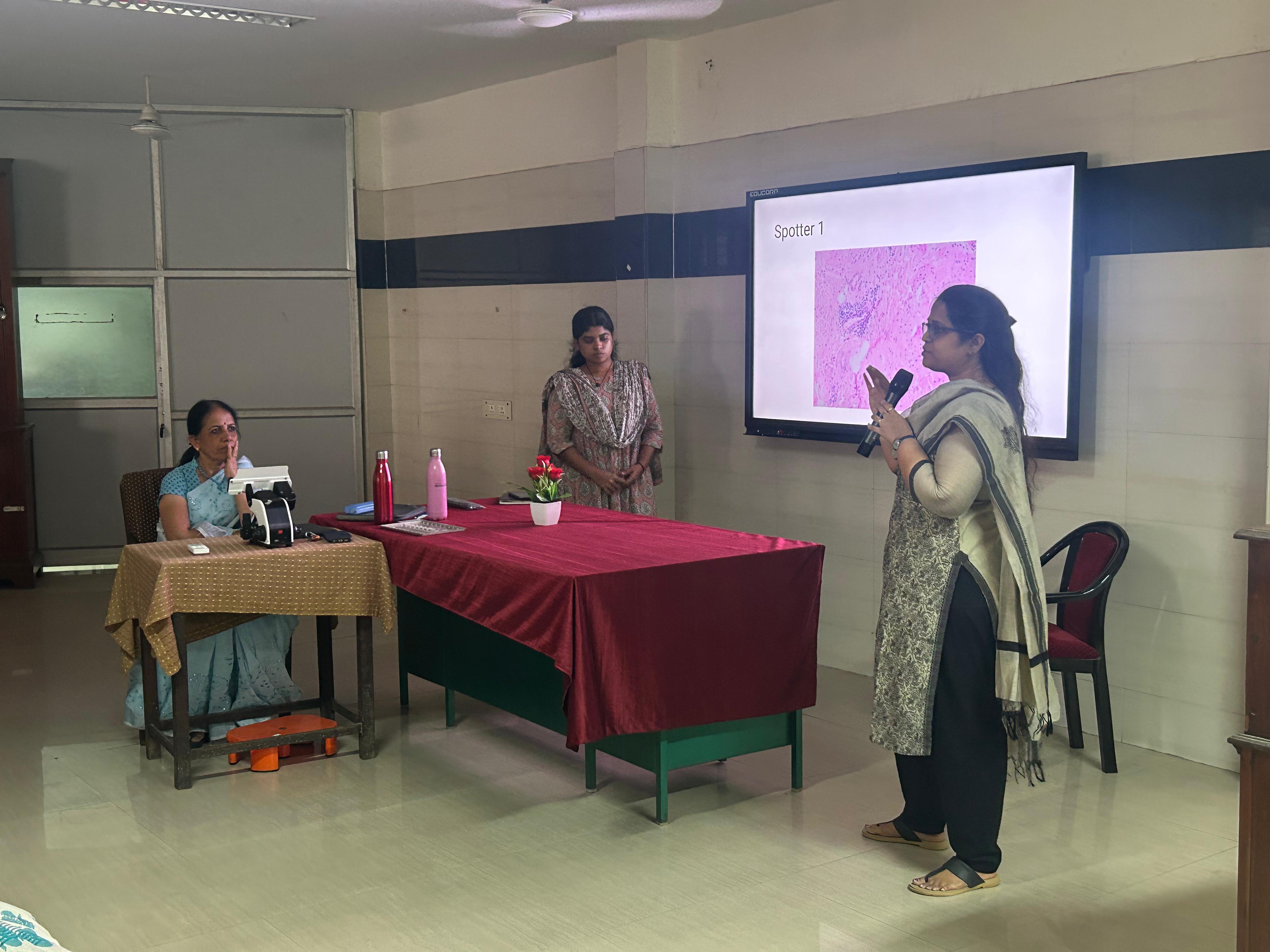
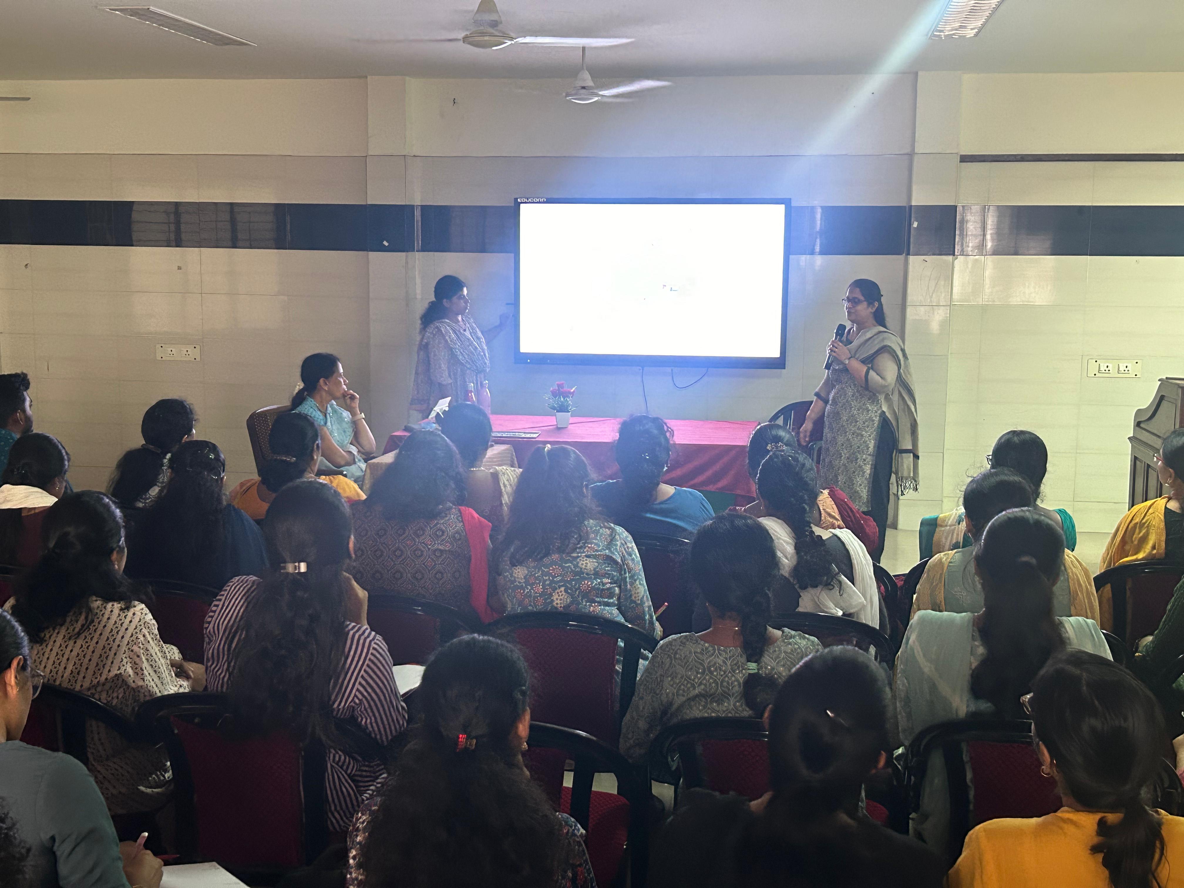
May 2022: Histopathology techniques workshop held.
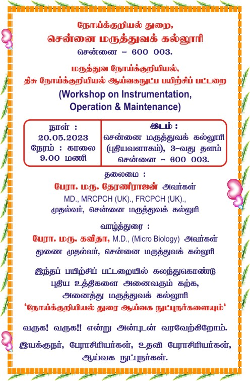
May 2023: Workshop on instrumentation, operation, and maintenance.
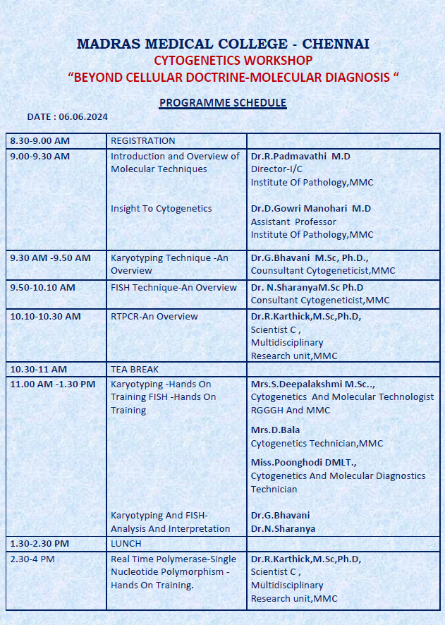
Sep 21, 2023: First Cytogenetics workshop (first among TN government colleges).
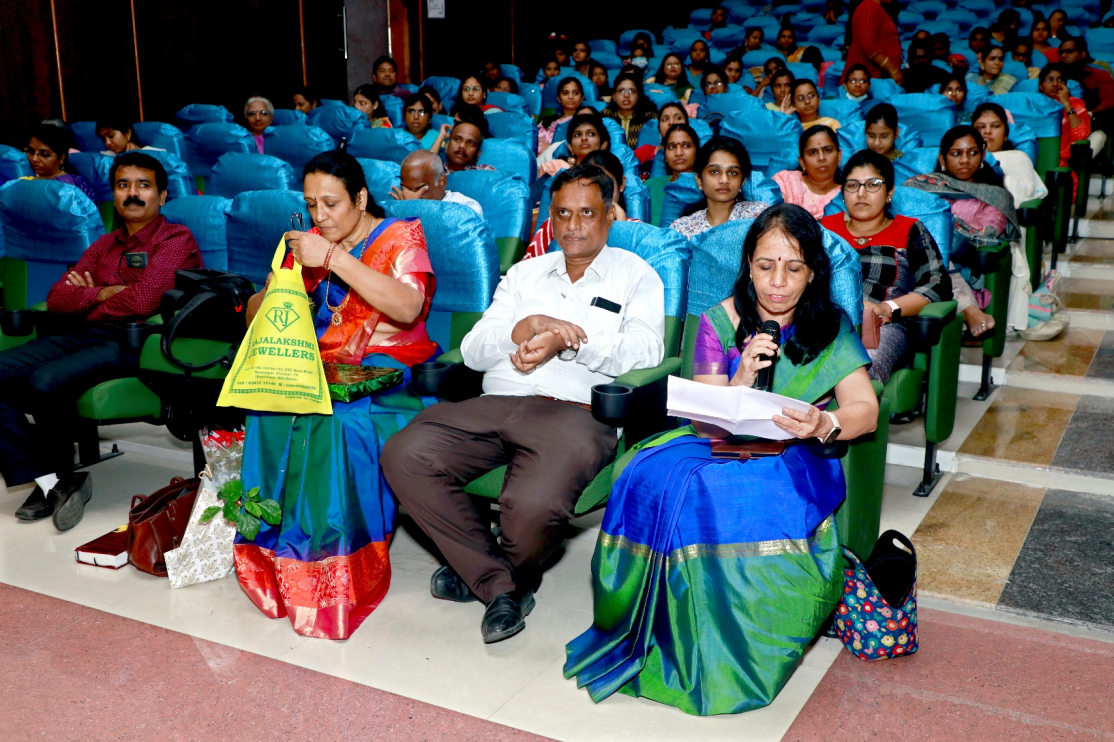
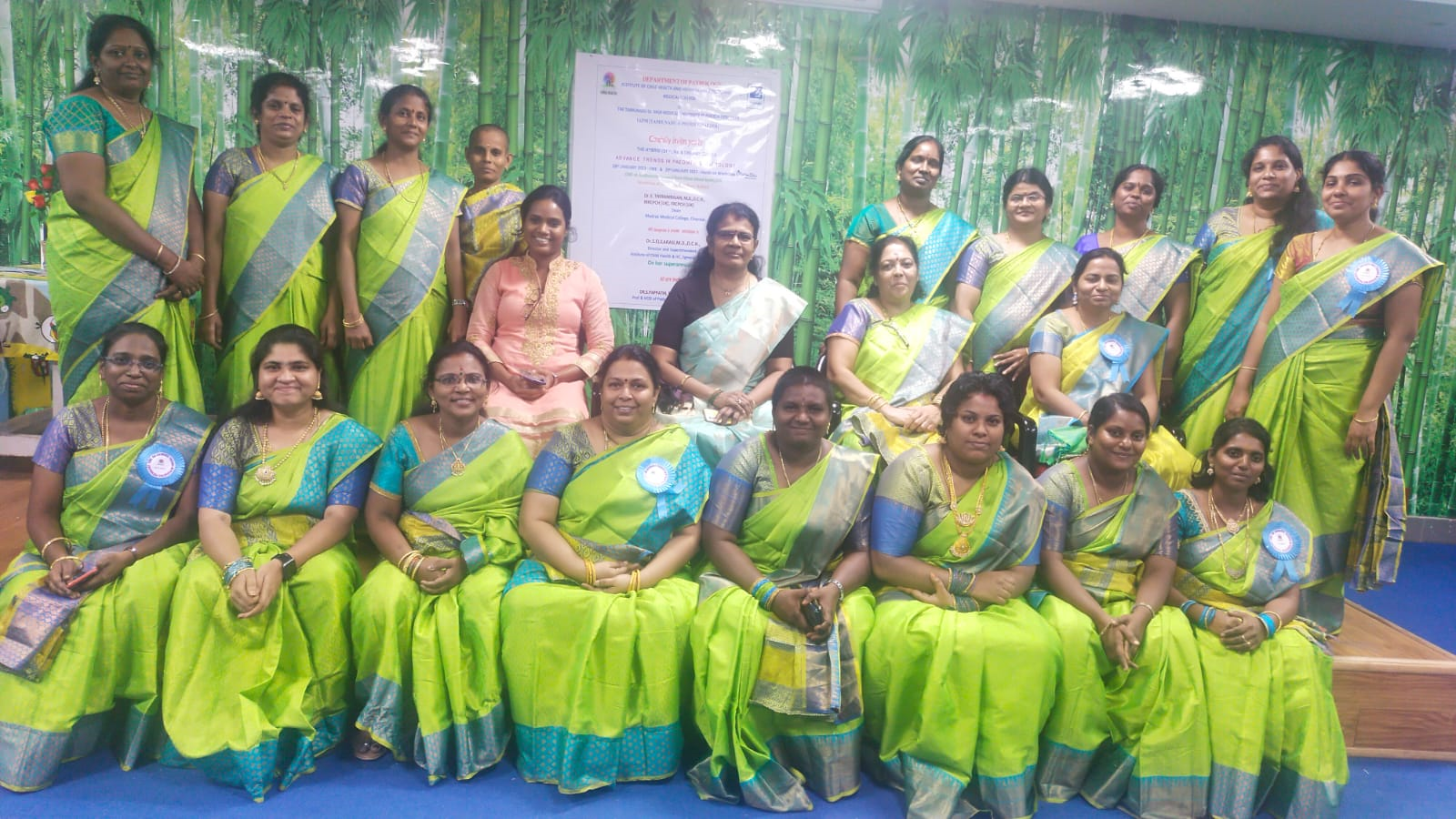
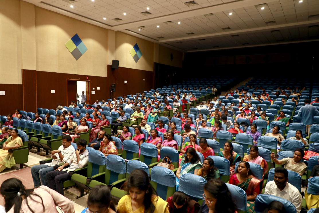
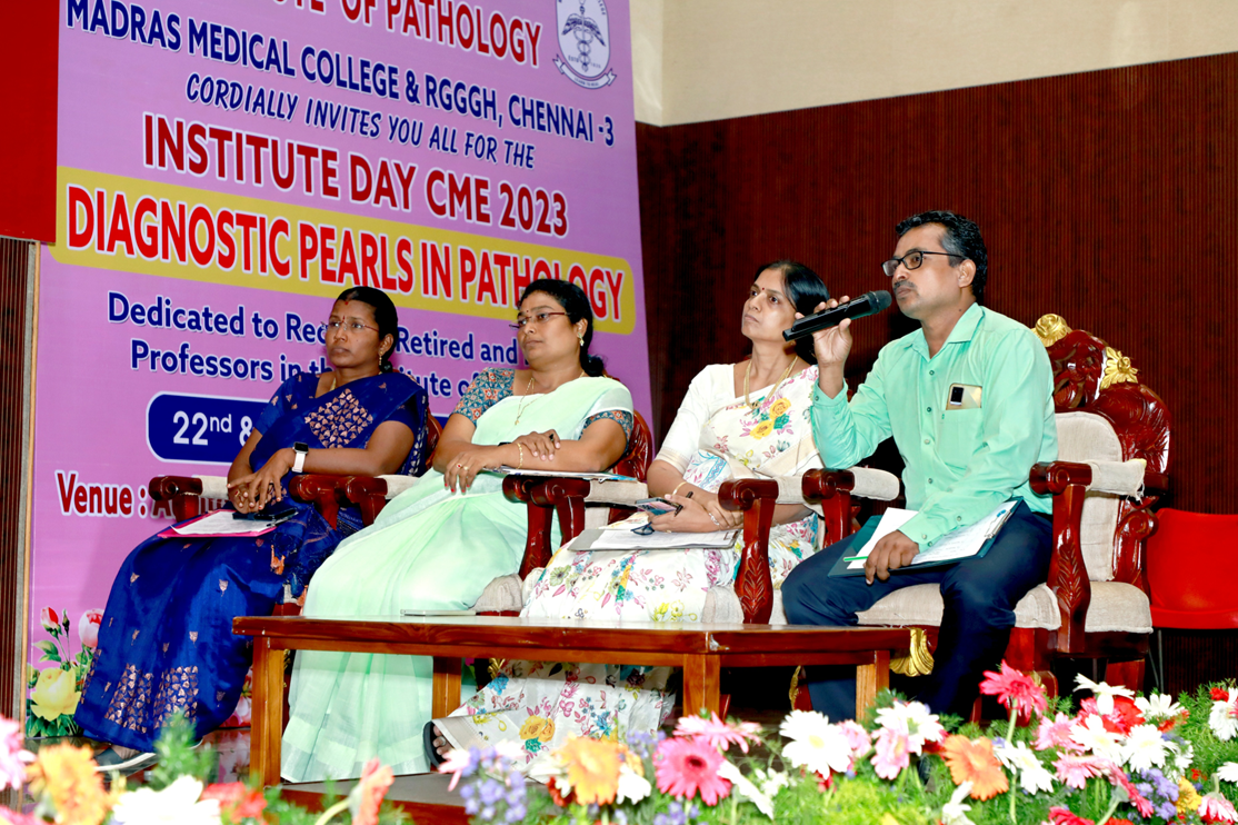
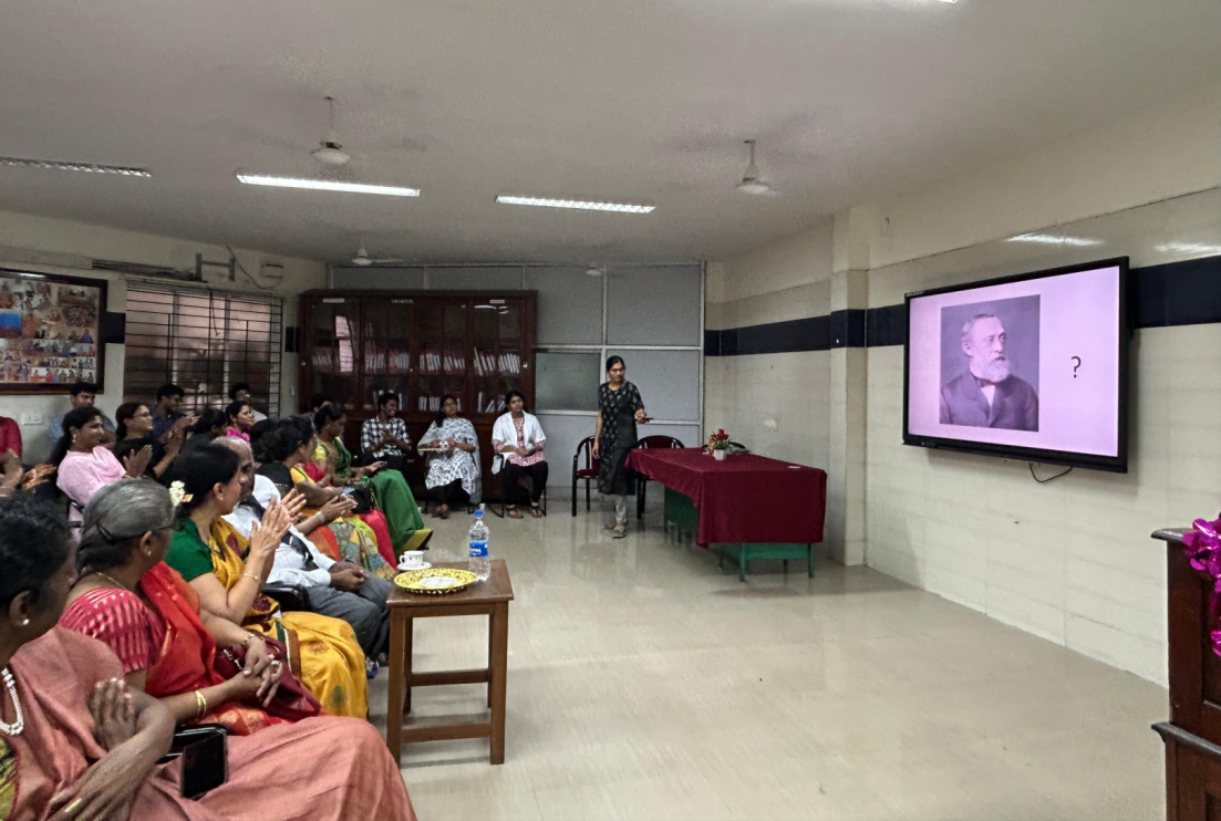
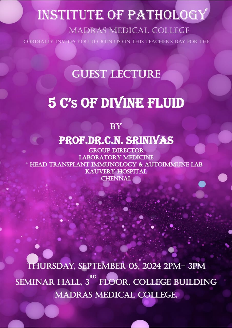
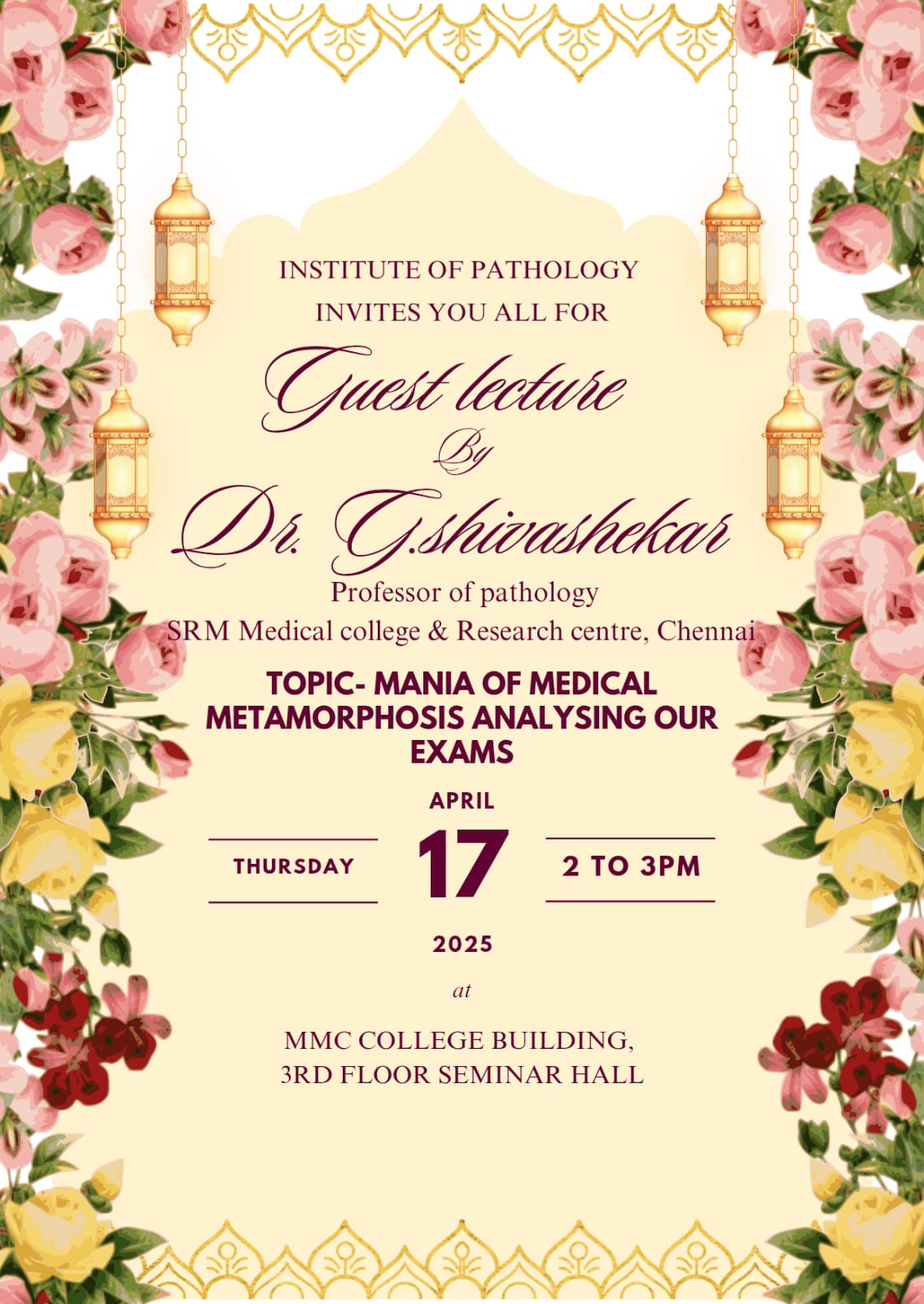
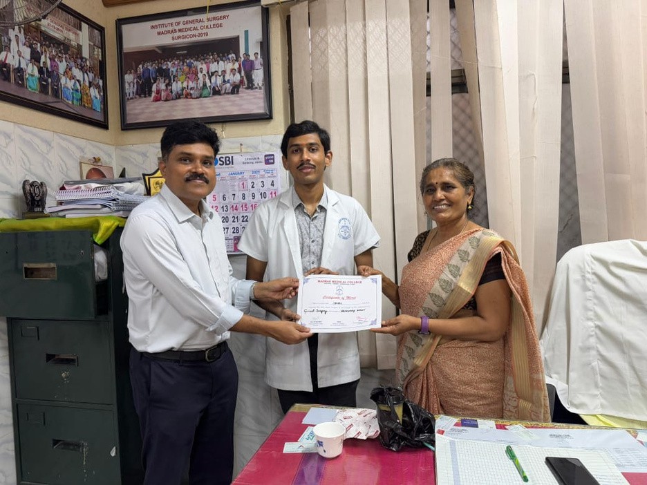
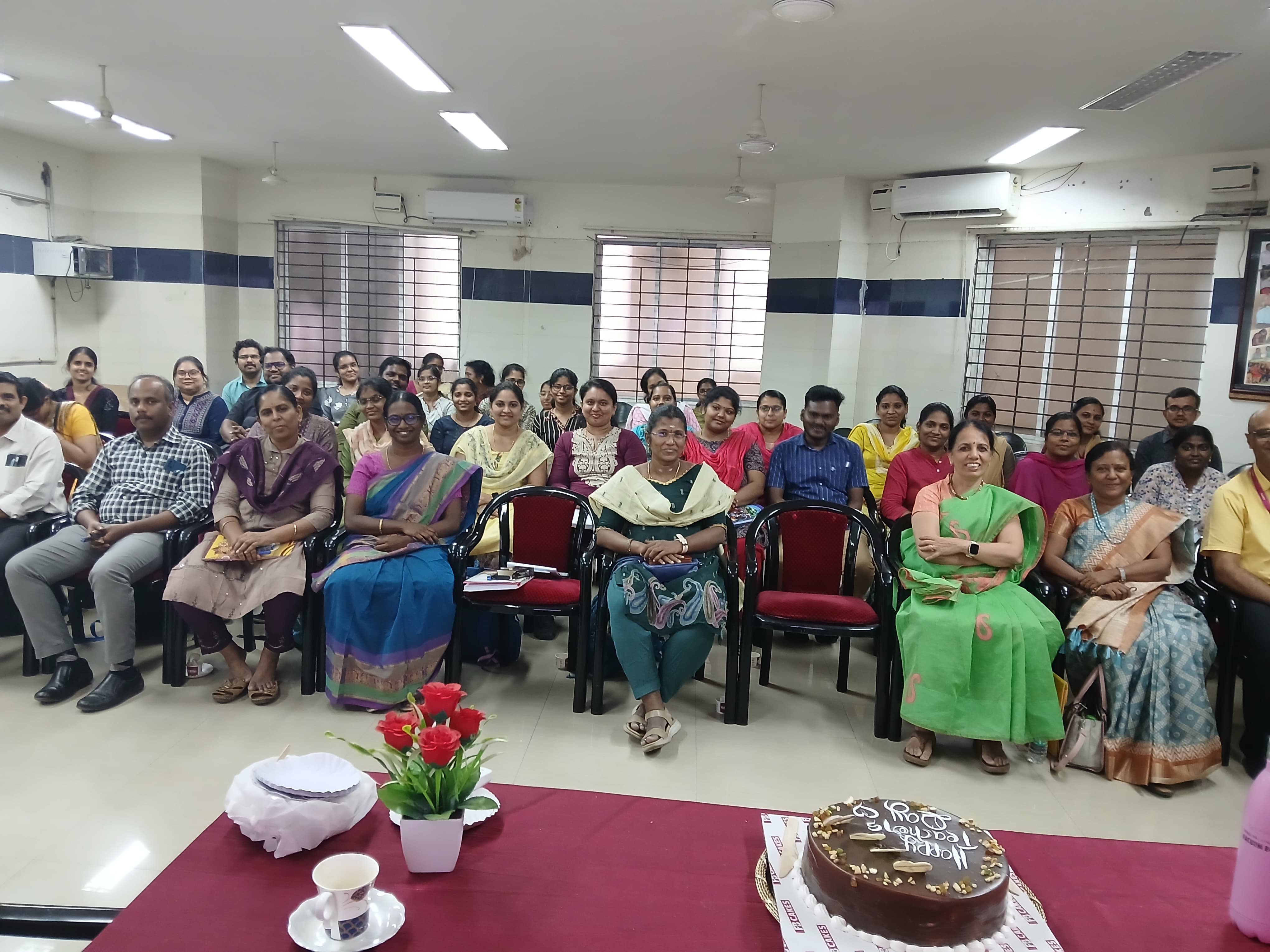
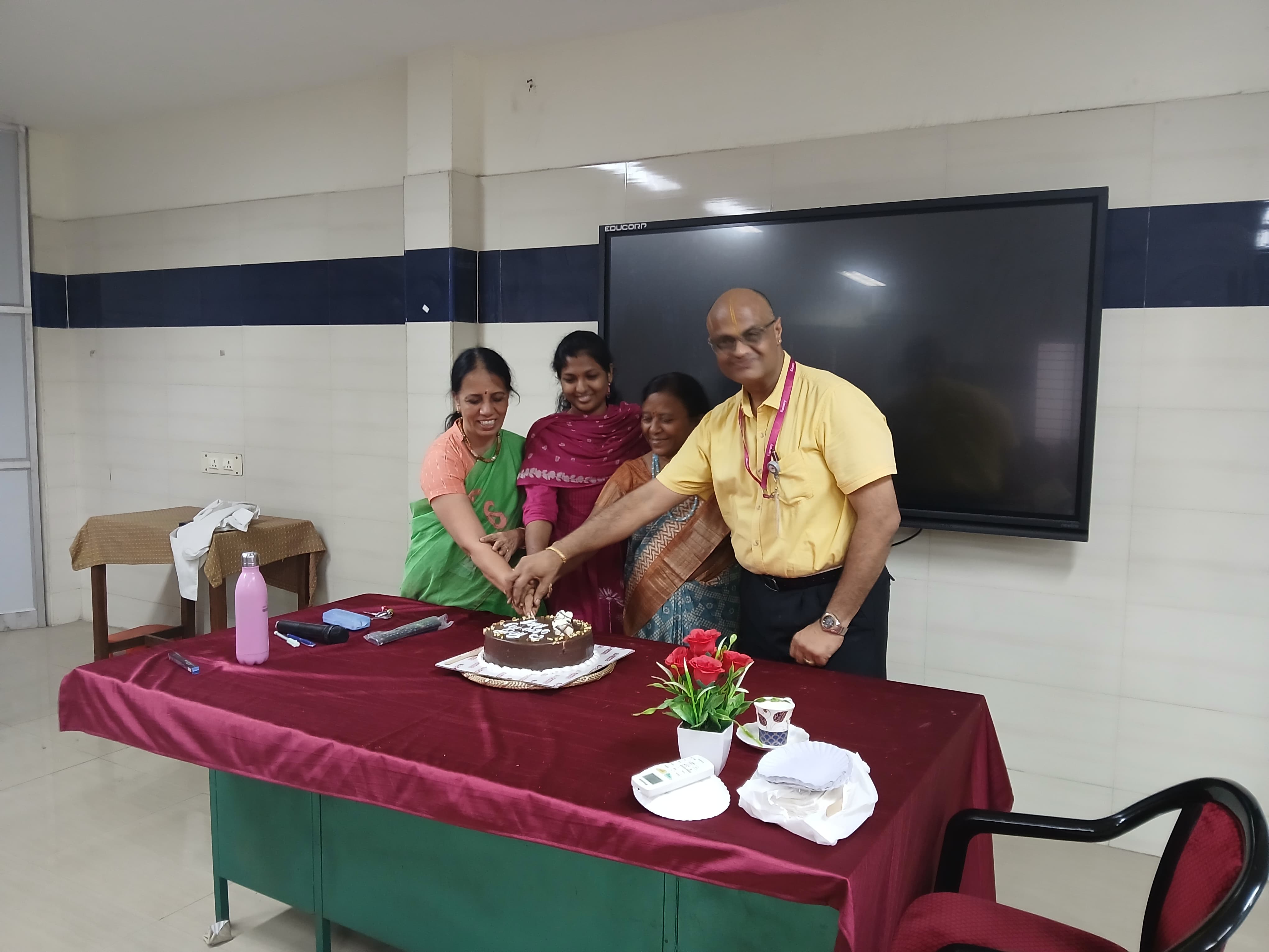
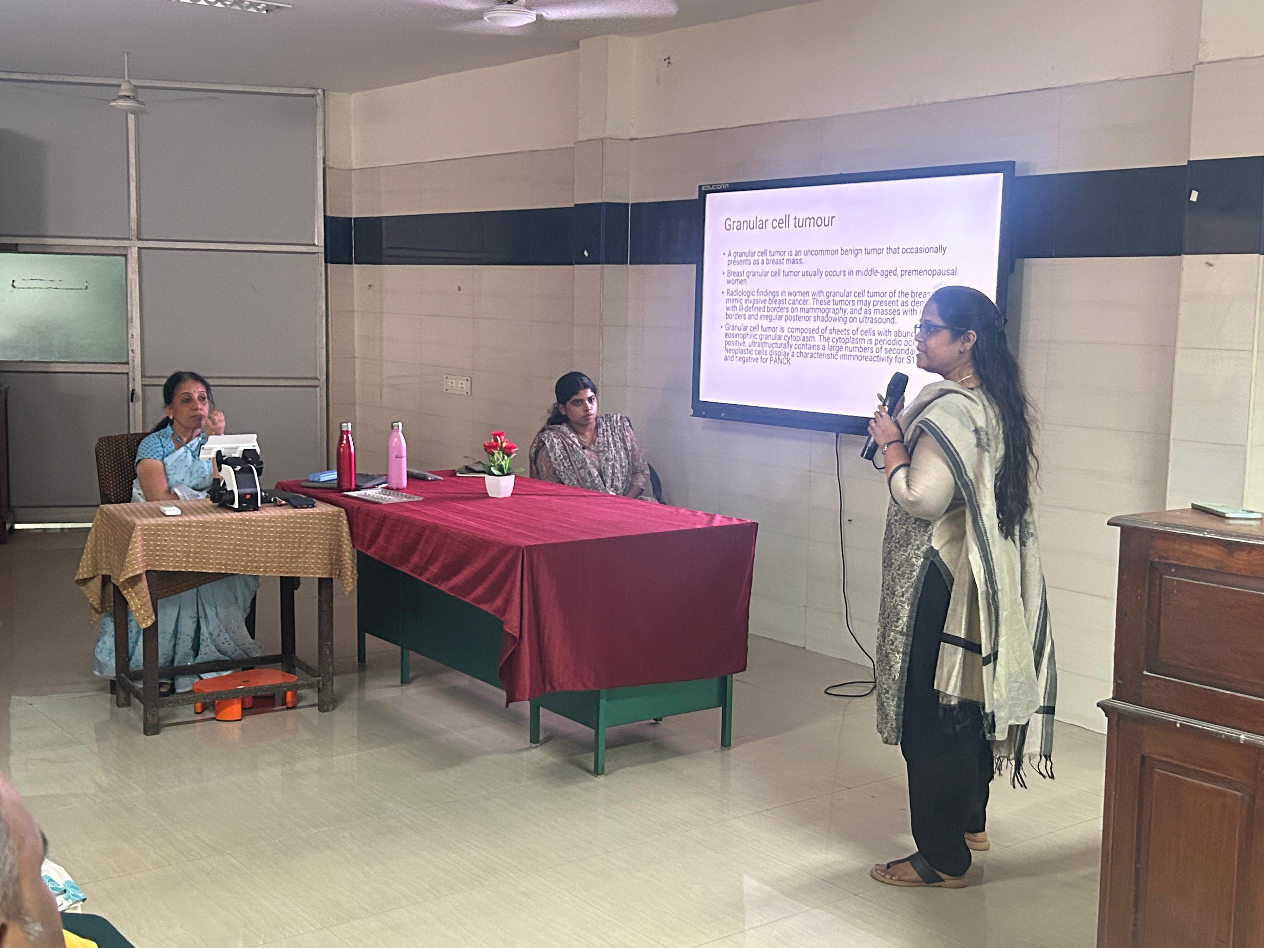
Sep 22–23, 2023: CME – “Diagnostic Pearls in Pathology” honoring retired and retiring professors.
Liquefaction time, Volume, Appearance, Colour, Viscosity, pH, Total sperm
concentration, Total motility, Vitality, Agglutination, Pus cells, RBCs,
Epithelial cells, Morphology
pH Paper / Naked eye examination / Morphology analyzed by manual pap stain
and microscopy
Stool
Component / Parameter
Method Used
Occult blood
Peroxidase Method
Urine (General Tests)
Component / Parameter
Method Used
Bile salt
Hay's test
Urine Bilirubin
Coupling with reagent / Manual-Smith test
Urine Blood
Peroxidase
Urine Glucose
Glucose Oxidase Peroxidase
Urine Ketones
Nitroprusside / Legal’s test
Urine Leucocytes
Esterase Activity of Granulocyte
Urine Nitrite
Griess test
Urine pH
Double indicator principle
Urine Protein
Protein error of pH Indicators / Sulphosalicylic acid Method [Manual]
Urine Specific Gravity
Ion exchange
Urine (Microscopy)
Component / Parameter
Method Used
Urine deposit: RBCs, Pus cells, Epithelial cells, Casts, Crystals, Organisms
Microscopy
Urine Urobilinogen
Coupling of urobilinogen with reagent
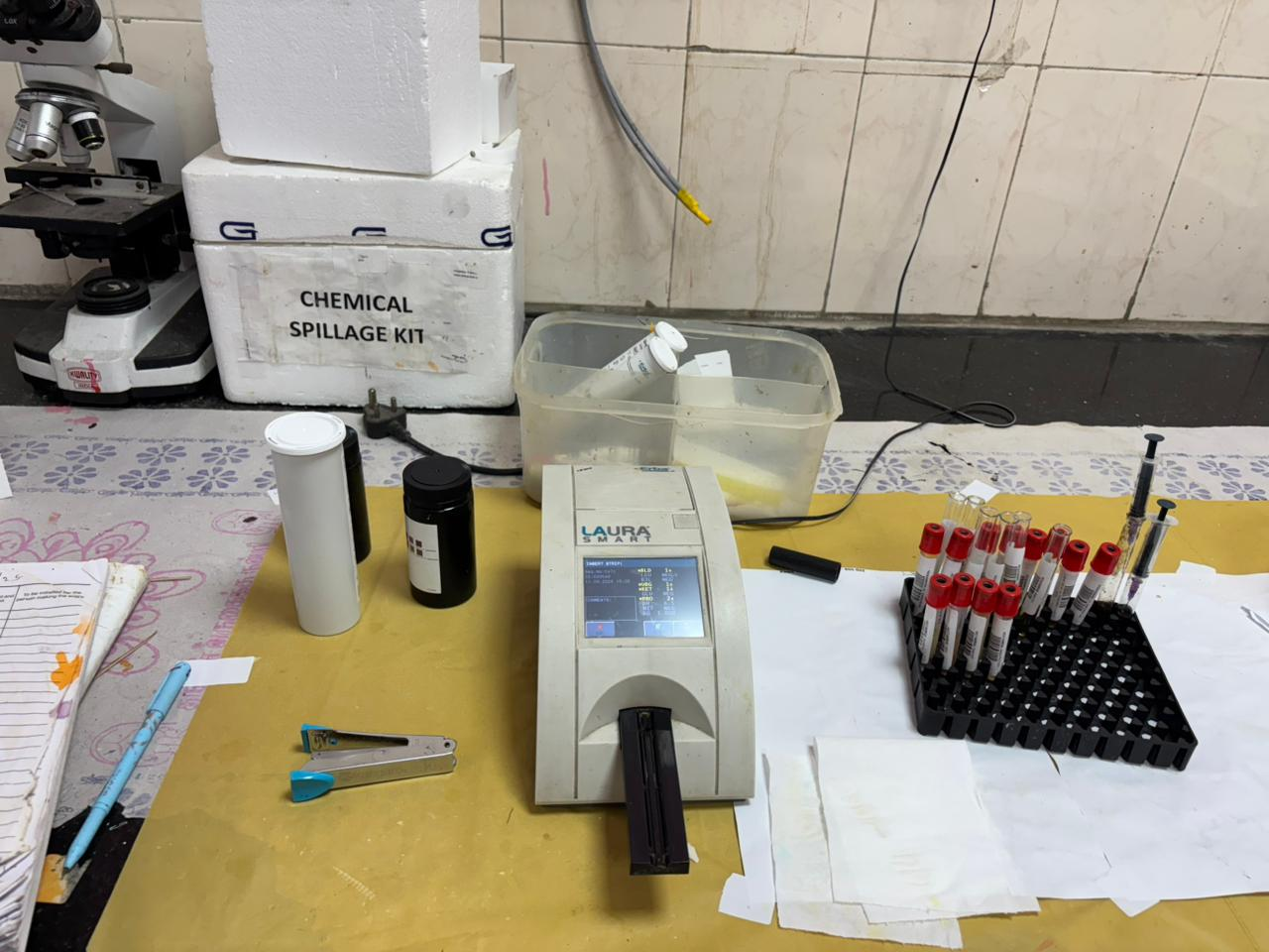
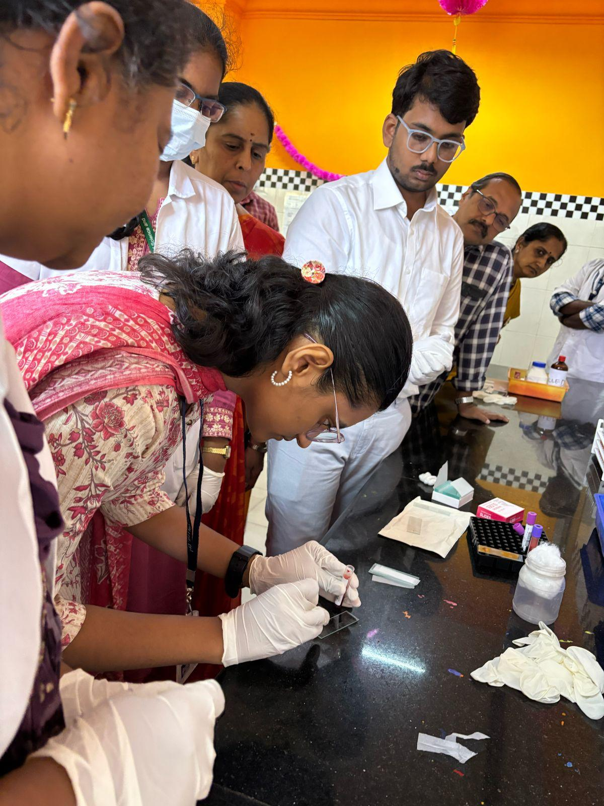
Bone Marrow Aspirate
Component / Parameter
Method Used
Bone Marrow Aspiration
Leishman Stain & Perl's Stain
Citrated Blood
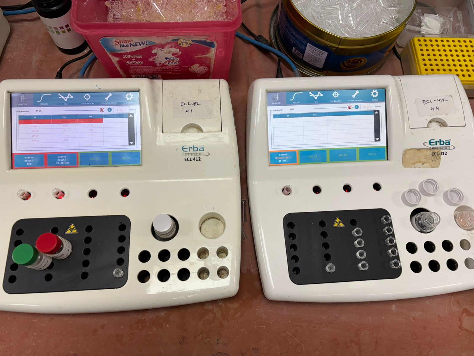
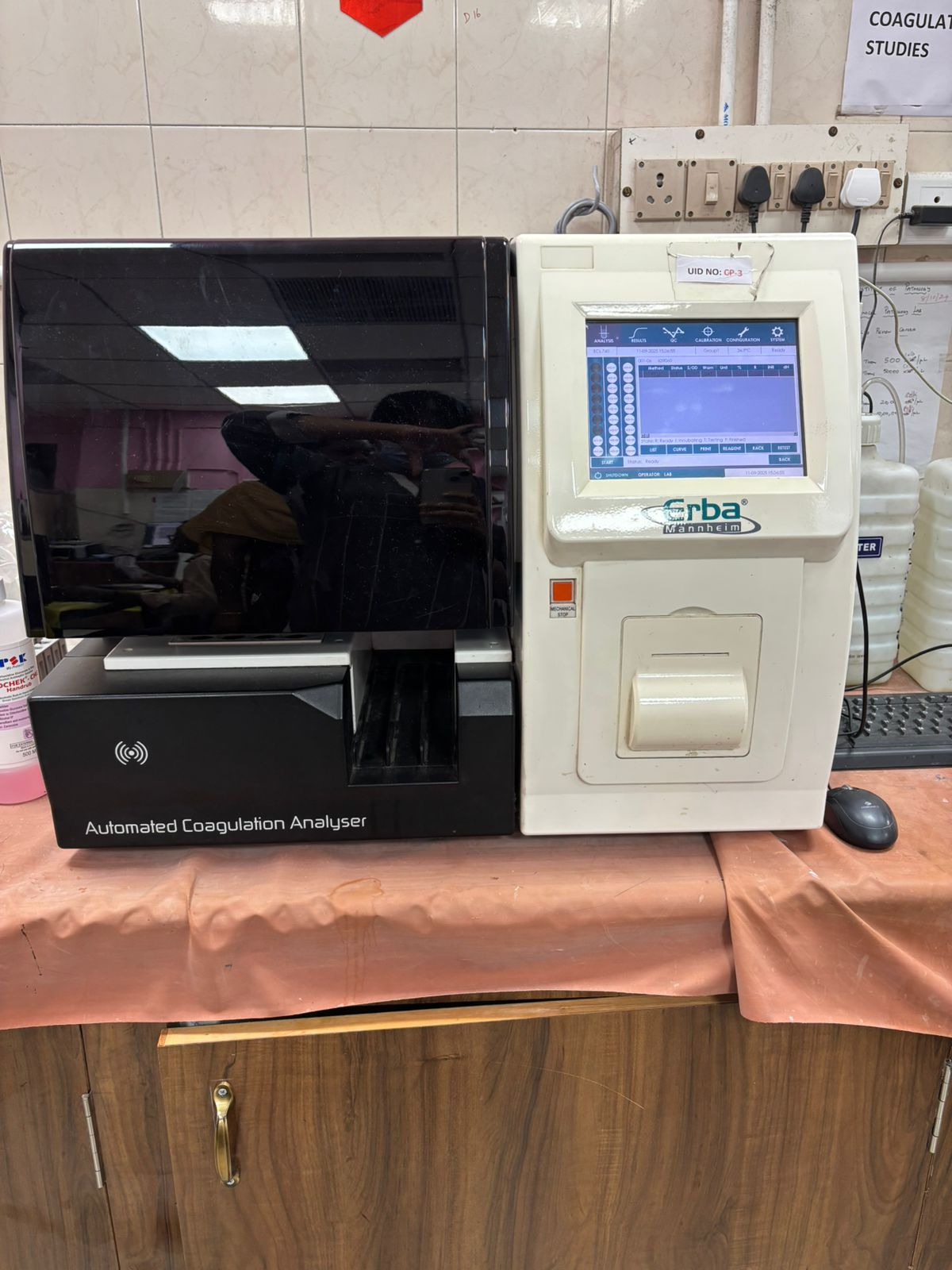
aPTT
Clot Based Optical Measurement
PT
Clot Based Optical Measurement
K2 EDTA Whole Blood
Component / Parameter
Method Used
Direct Coomb's test
Agglutination of antihuman globulin
Hematocrit
RBC Pulse Height Detection Method
Hemoglobin
SLS Hemoglobin Method
MCH
Calculation with Hemoglobin / RBC
MCHC
Calculation with Hemoglobin / Hematocrit
MCV
Calculation with Hematocrit / RBC
Osmotic fragility test
Lysis by hypotonic saline / Manual observation
PAS stain, Myeloperioxidase stain, Sudan black, Non specific esterase
Staining and microscopy
Peripheral Smear
Leishman Stain
Platelet
Hydrodynamic Focussing (DC Detection)
RBC
Hydrodynamic Focussing (DC Detection)
Reticulocyte count
Fluorescence flowcytometry
Reticulocyte count
Manual using New Methylene Blue stain and microscopy
Sickle cell preparation
Microscopy / Metabisulphite preparation
Smear for malarial parasite
Thin smear - Leishman stain and Thick smear - Field stain / Microscopy
Smear for microfilaria
Microscopy / Wet mount preparation / Leishman stain