SUCCESS STORY 1:
Tracheal Resection and Cricotracheal
Anastomosis:
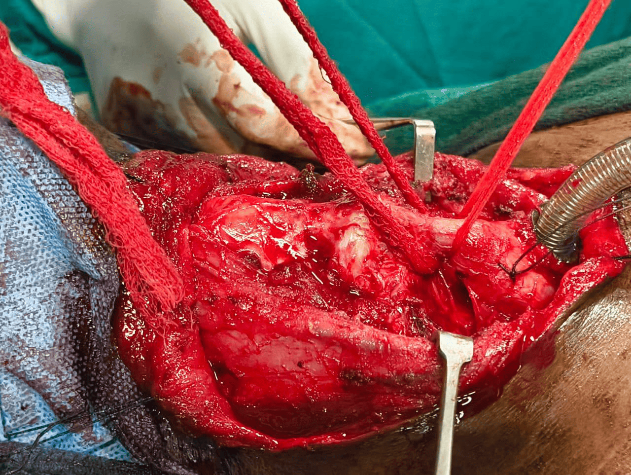
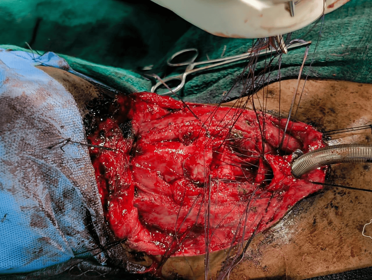
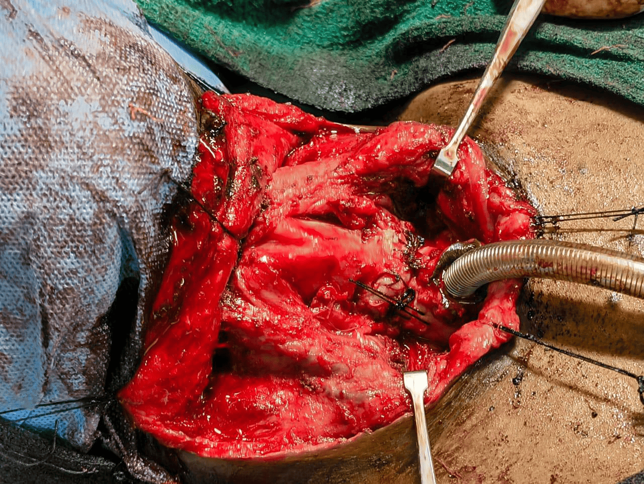
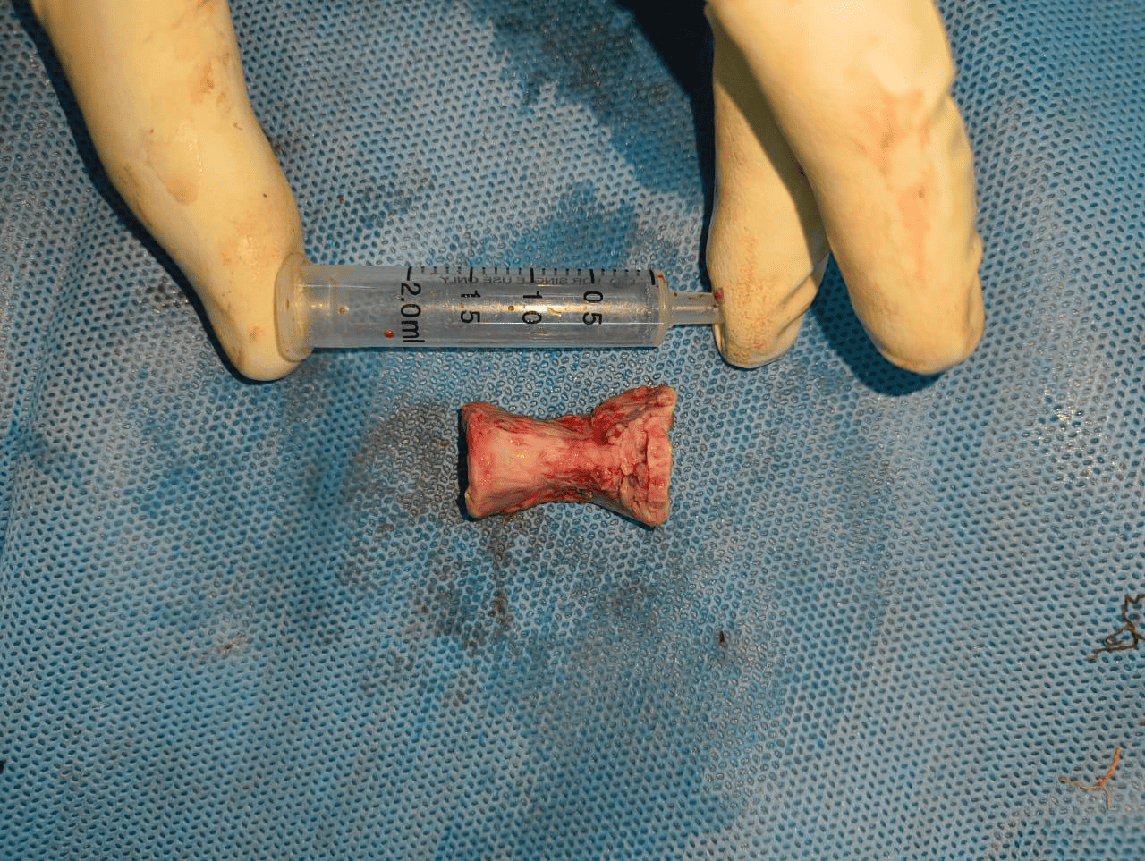
At our institute, we successfully managed a challenging case of severe
post-intubation subglottic stenosis in a 31-year-old male who had a history of
prolonged ventilation following a suicide attempt. The patient had previously
undergone multiple failed procedures including dilatations, laser treatments,
and micro-laryngeal excision elsewhere. At our center, he underwent definitive
surgical management with tracheal resection and cricotracheal anastomosis,
planned using detailed 3D CT imaging. Postoperative care included staged
bronchoscopy, nutritional management, and airway surveillance. This case
highlights the advanced airway reconstructive capabilities at our institution
and underscores the importance of a multidisciplinary approach in managing
complex airway stenosis.
SUCCESS STORY 2:
Recurrent Juvenile Nasopharyngeal
Angiofibroma with intracranial extension:
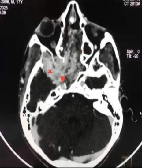
17Years/ male, presenting with torrential epistaxis. He is a known
case of Juvenile Nasopharyngeal Angiofibroma operated twice before. JNA surgery
is considered as one of the most difficult of surgeries in Skull base surgery
being a vascular tumour and revision JNA surgery requires the highest-level
skill set. His imaging showed an extensive recurrence. It was a fibrous
adherent tumour completely occupying both nasal cavity, nasopharynx, right PPF,
Infratemporal Fossa, Middle cranial fossa, Cavernous sinus and 270-degree encasement
of right paraclival carotid. After having a multidisciplinary discussion with
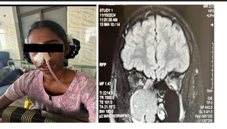
Neurosurgeon, Interventional radiologist and after embolising the tumour the
patient was taken up for surgery. It was a six hours long surgery and we
successfully dissected the tumour off the middle cranial fossa, cavernous sinus
and para-clival carotid and performed a complete excision of the tumour. The
patient has clinically improved and is under constant follow up.
SUCCESS STORY 3:
Cochlear Re-implantation:
UIORL is a referral center for complex
cochlear re-implantation surgeries, offering renewed hearing hope to patients
with failed or non-functional implants. Re-implantation is a technically
demanding procedure requiring precise surgical planning, detailed radiological
evaluation, and experience in managing fibrotic beds and electrode damage. The
center is equipped with dedicated audiological testing and rehabilitation
facilities, allowing for seamless post-operative integration and speech
therapy. This has been a transformative service for hearing-impaired children
and adults across Tamil Nadu and other states.
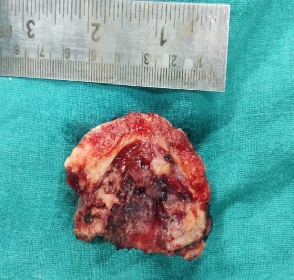
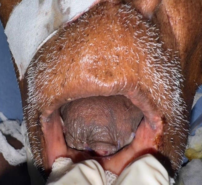
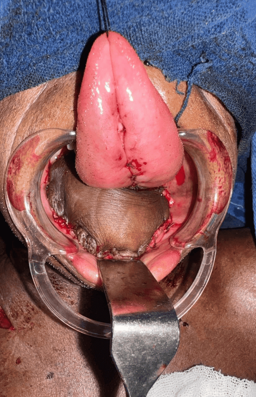
SUCCESS STORY 4:
Angiomatous nasal polyp:
A 60-year-old female came with complaints of right sided nasal
obstruction and nasal discharge for 6 months with loss of smell sensation. CECT
PNS revealed an enhancing vascular mass in the right nasal cavity extending to
the nasopharynx and CT Angiogram showed feeding vessel from the ascending
pharyngeal artery. Preoperative embolization of the feeding vessels was done
and the patient underwent endoscopic excision of the nasal mass which was sent
for histopathological examination; results showed the mass to be an angiomatous
variant of antrochoanal polyp. Postoperative period was uneventful and patient
recovered well
SUCCESS STORY 5:
Dentigerous cyst:
A 11-year-old male child Came with Complaint of Right-side facial
swelling for 1 year swelling (+) Right side of cheek 3× 2cm. Skin over swelling
_ normal. Swelling extend medially from the middle 1/3 rd dorsum of nose &
1cm below lateral canthus – about 3×2cm oval in shape. Firm in consistency skin
above the swelling normal. CT facial bone: - Soft dense lesion noted in
superior alveolar margin extending up to Right maxillary sinus. Right DENTIGEROUS
SWELLING. Procedure done: - endonasal endoscopic dentigerous cyst excision done
via inferior meatal antrostomy with complete removal of the unerupted tooth
lying in the maxillary sinus cavity
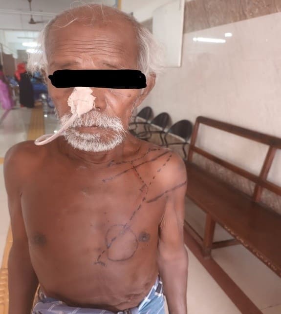
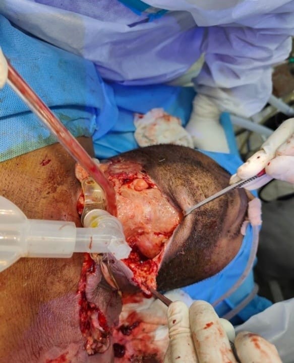
SUCCESS STORY 6:
Bilateral neck dissection with
composite resection (Marginal mandibulectomy with wide local resection) with
Pectoralis Major Myocutaneous Flap Reconstruction:
A 70 year male, came with complaints of swelling in the floor of mouth since
1month. The patient had swelling in the left level IB lymph node level. The
patient had difficulty in protruding the tongue and swallowing and hence was on
Ryle’s tube feed. Biopsy and FNAC revealed the diagnosis of Adenocarcinoma of
floor of mouth. The tumour was staged as Stage IVA on further radiological and
clinical assessment. The case was taken up for discussion in tumour board and
it was decided to perform surgery followed by adjuvant radiation. A
multidisciplinary team including ENT, OMFS, plastic surgery and anaesthesia
were involved in the procedure. The ENT team performed a left Anterolateral
comprehensive neck dissection and right Supraomohyoid neck dissection, then the
ENT and OMFS team performed a composite resection of the tumour (extended
Marginal mandibulectomy and wide local resection). A tracheostomy was performed
for maintaining the airway. Then the plastic surgery team took over and
performed the reconstruction with a Pectoralis Major Myocutaneous flap (PMMC).
The patient was later managed in the ICU and after 3 weeks patient was started
on oral feeds after performing a Functional endoscopic evaluation of swallowing
and was successfully weaned off the tracheostomy. Histopathology was Mucinous
adenocarcinoma with left level IB, II and III lymph node positivity. The patient is due for
postoperative radiotherapy.
SUCCESS STORY 7:
Advanced Juvenile Nasopharyngeal
Angiofibroma with Intracranial Extension
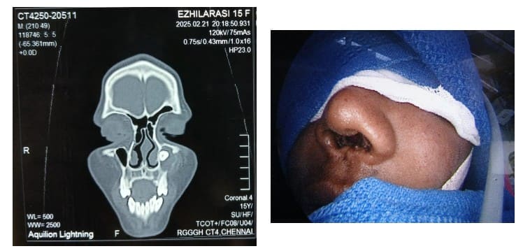
A 15-year-old male
from Trichy presented with a 2-year history of right facial swelling, bilateral
nasal obstruction for the past year, nasal discharge, mouth breathing, and
snoring. Radiological evaluation showed a Radowksi Stage 3B Juvenile
Nasopharyngeal Angiofibroma with evidence of intracranial extension into the
middle cranial fossa without Dural breach. Preoperative embolization of the
right internal maxillary and ascending pharyngeal arteries was performed,
followed by surgical excision of the tumour via the maxillary swing approach
for optimal access and control

SUCCESS STORY 8:
Pituitary thyrotropinoma:
A
28year old female, found to have hyperthyroidism during routine TFT at the time
of pregnancy and on antithyroid drugs, presented with palpitations, excessive
sweating, oligomenorrhea, tremors and neck swelling. On examination, a neck
swelling of size 7x8cm in the anterior aspect of the neck was noted. TSH was
found to be 72 microIU/ml and FT4 was 3.2 ng/dl. Radiological investigation
were as follows: USG neck: p/o MNG Right lobe-3.8*2.2*3.8cm Left lobe
-2.5*1.7*3.9cm CT PNS -e/o well defined non enhancing hypodense lesion noted in
pituitary region measuring 9.2mm MRI brain with Sella contrast: Well-defined T2
heterogeneous lesion in right side of pituitary gland -hyper enhancing in post
contrast region MRI brain - no significant abnormalities Patient was diagnosed
as Pituitary macroadenoma (thyrotropinoma) and proceeded with endoscopic trans
nasal excision of the mass. Histopathology showed it was a pituitary
neuroendocrine tumour. Postoperative hormone profile was done including TFT
which showed TSH was on the decreasing trend. Now the patient is stable and on
regular follow up.
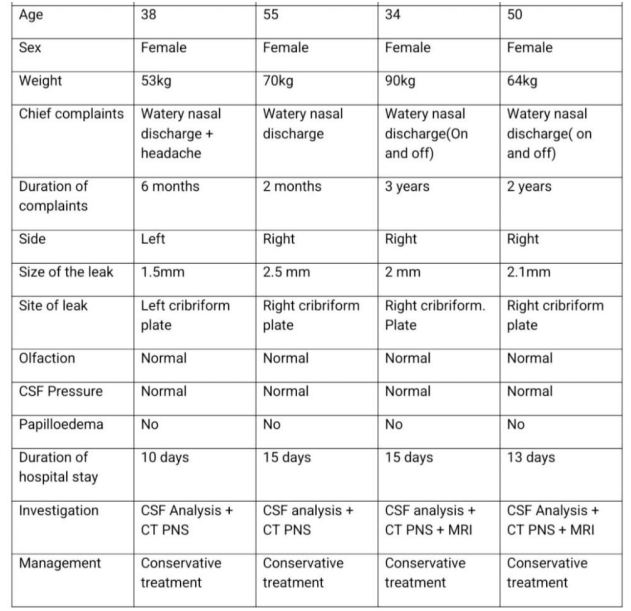
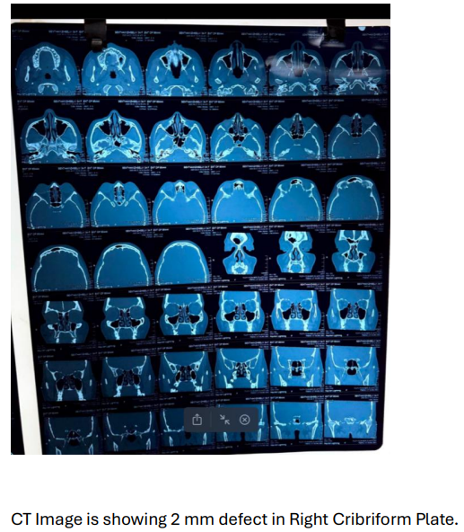
SUCCESS STORY 9:
Spontaneous CSF rhinorrhea: - In this study, patients from age group of 50-60 years old female
had complaints of watery nasal discharge and headache, on and off on right side
for a period of 2 years. Patients had defect of size 2-2.5 mm on right
cribriform plate. Patient had no evidence of papilledema. Patients had no fluctuation
of ICT. Patients managed conservatively. Another age group of patients from
30-40 years females came with complaints of watery nasal discharge and headache
on both right and left nasal cavity with defect of size 2-2.5 mm of both right
and left cribriform plate. Patients has no evidence of papilledema. Patients
managed conservatively.
Patients were managed with Tablet Acetazolamide
250mg 2tds.Till the CSF RHINORRHOEA completely stopped. Then the dosage reduced
to 1tds & continued for 3-month period. Patient reviewed om monthly
interval for a period of 2 year. To see the any recurrence of the disease. CSF
leak from the Anterior skull base which are SPONTANEOUS in nature & most
common in female can be successfully managed conservatively. Surgical intervention
needed if the conservative therapy fails, or associated with meningocele or if
there is any suspicion of intracranial complications.
SUCCESS STORY 10:
Traumatic Optic nerve
decompression:
A case of post-traumatic optic neuropathy in a
young male who presented with acute vision loss in the right eye following
facial trauma. After thorough evaluation and imaging, the patient was diagnosed
with compressive optic neuropathy. A trans nasal endoscopic optic nerve
decompression was performed to relieve pressure on the optic nerve.
Postoperatively, the patient showed remarkable visual improvement – progressing
from only perception of light to being able to count fingers close to the face.
SUCCESS STORY 11:
Diffuse large B cell lymphoma of
thyroid: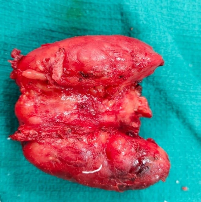
A 77-year-old female came to our OPD with
complaints of a huge neck swelling in front of the neck for the past 2 months.
On doing radiological investigations the thyroid gland was found causing mass
effect in the form of compression of trachea of minimum diameter 9.6mm
(transverse). The patient was operated
and total thyroidectomy done. A highly challenging surgery and the thyroid
gland with isthmus was removed in toto. The specimen was sent for
histopathological examination which revealed diffuse large B cell lymphoma.
Tumour board opinion was obtained for the same and patient followed up in
medical oncology ward with RCVP regimen.
SUCCESS STORY 12:
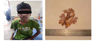
Foreign body removal:
A 95-year-old hypertensive female presented to the casualty with
alleged history of foreign body ingestion (dentures) at around 11:00 followed
by pain during swallowing and foreign body sensation in the throat. Video
laryngoscopic examination revealed mucosal injuries and oedema in the
vallecula, possibly due to induced vomiting, and the structures further down
could not be visualized due to pooling of saliva. X-ray lateral view of the
neck revealed the presence of large dentures at the level of the cricopharynx.
Patient also had scoliosis and thrombocytopenia in addition to hypertension.
Emergency anaesthesia fitness was obtained and the patient was immediately
taken up for rigid esophagoscopy and foreign body removal. The denture was
removed and the patient was observed postoperatively in the ICU. No
complications were encountered, the patient recovered well and was discharged.
SUCCESS STORY 13:
LEFT COM / CSF OTORHOEA / LABYRINTHITIS:
A
24-year-old male patient came with complaints of severe headache for 4 days,
frontal region, left ear discharge for 6 months, on local examination, left ear
minimal discharge, pulsatile watery discharge seen deep inside the middle ear.
On further radiological imaging, Fungus cerebri (7*2cm) noted. Intracranial
abscess managed conservatively with neuro surgery opinion foe 3 weeks with
improvement in clinical condition. Patient taken up for left revision mastoidectomy
with blind sac closure and reconstruction of Tegmen defect. Patient was
discharged after a month with complete resolution of intracranial
complications.
SUCCESS STORY 14:
Recurrent CSF Rhinorrhoea:
A 41year female from Vellore came with complaints of watery nasal
discharge from the left nasal cavity for the past 20 days. She had a previous history of undergoing CSF
leak repair in a private hospital in Vellore. On undertaking various MRI
Cisternogram revealed bilateral CSF leak with left anterior encephalocele.
Intraoperatively, a defect of size 1cm*0.5 cm seen in left cribriform plate.
Defect closed in layers with first layer surgical, fascia lata kept as second
layer, hadad flap as third layer and gel foam as final layer, making sure not
to obstruct frontal and sphenoid sinus opening. The patient was managed post-
operatively with anti-epileptics and T. Acetazolamide. The procedure was
followed by a positive outcome and the patient has been in follow up for
regular CSF pressure checking and placement of lumbar drain if required in view
of benign intracranial hypertension
SUCCESS STORY 15:
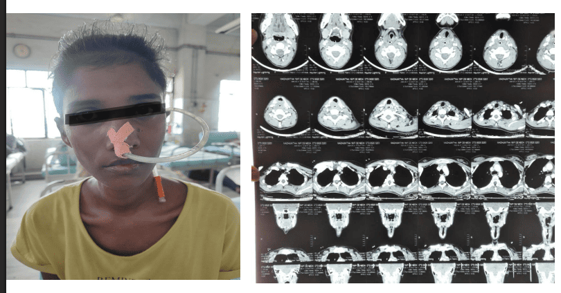
Foreign
body removal surgeries in airway: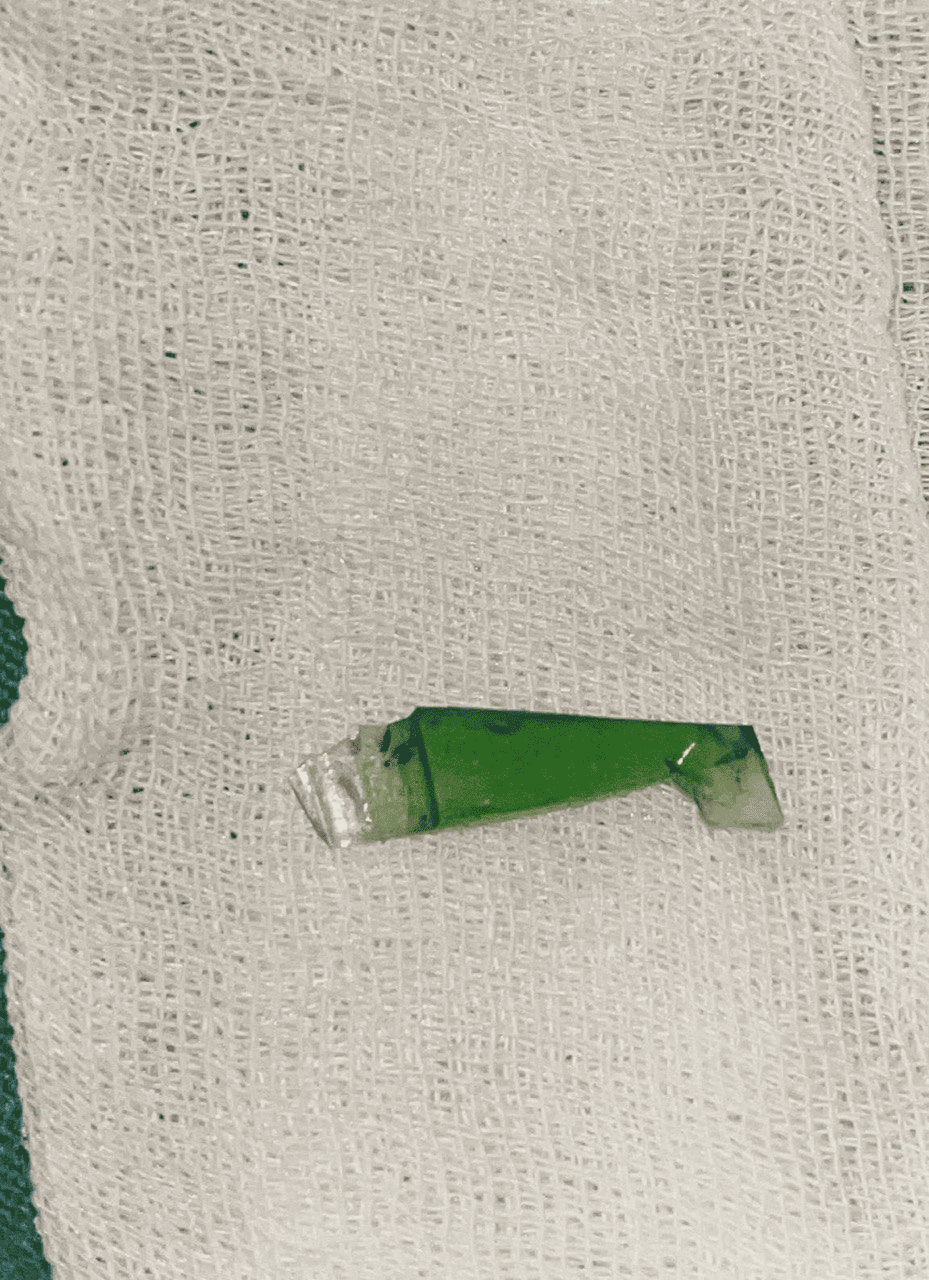
A 27-year-old male aspirated a glass foreign
body into the right lower lobe bronchus. In view of the foreign body's
distal location in the right lower lobe bronchus and limited reach using
conventional optical forceps, an innovative hybrid bronchoscopic technique was
employed. A flexible bronchoscope was secured with microlaryngeal forceps and
inserted through the barrel of a rigid bronchoscope. This hybrid approach
allowed enhanced maneuverability and visualization, better angulation, and
successful retrieval of the foreign body- glass piece with minimal mucosal
trauma. The technique proved to be a practical and effective alternative in
situations where conventional tools may be inadequate or unavailable.
SUCCESS STORY 16:
Recurrent juvenile nasopharyngeal
angiofibroma:
A 14-year-old male presented with
complaints of bilateral nasal obstruction for 6 months with one episode of
epistaxis 6 months back. The patient was already diagnosed as a case of
juvenile nasopharyngeal angiofibroma in 2020 and 2022 and was operated for the
same twice in Vellore Medical College. At present. CECT PNS revealed an
enhancing mass in the entire nasopharynx with intracranial extension (Radkowski
stage IVB) and CT angiogram revealed feeding vessels from the Internal
Maxillary Artery. Preoperative embolization of the feeding vessels was done by
Interventional Radiology after which the patient underwent revision endoscopic
excision of the nasopharyngeal angiofibroma for the third time. Postoperative
period was uneventful and the patient was discharged.
SUCCESS STORY 17:
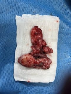
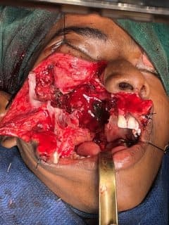
Ameloblastoma:
A 28 YEARS old female Patient came with Complaint of swelling
in the Right side of cheek in the last 3 year. Biopsy taken from the swelling
and proved as Desmoplastic ameloblastoma. Swelling (+) Right cheek measuring
5×4cm smooth, skin above the swelling normal. CT PNS: - Desmoplastic
ameloblastoma. Ill-defined expansive cystic lesion in Right maxilla. Procedure
done: - Right partial maxillectomy with obturator fixation. (sub labial
approach) HPE report: - Desmoplastic ameloblastoma.
SUCCESS STORY 18:
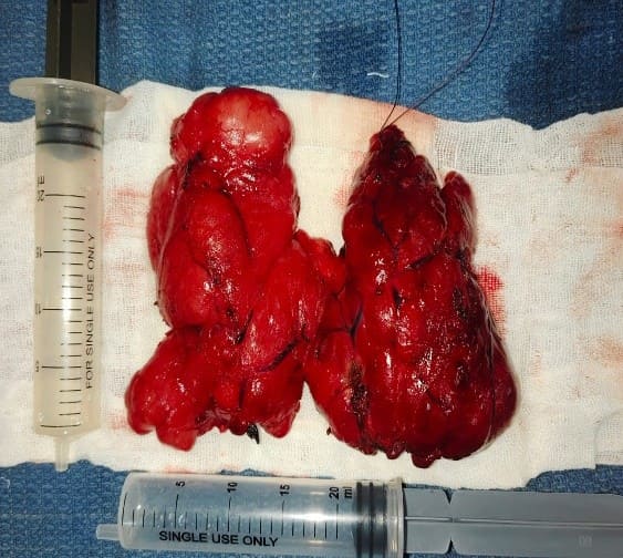
Multinodular goitre
thyroid with retrosternal extension:
61-year-old
female came to the OPD with complaints of swelling in front of the neck for the
past 3 years. Thyroid function test showed the patient in euthyroid status.
On further radiological examination revealed
the mass to be compressing the trachea with retrosternal extension of
approximately 2cm below thoracic inlet.
This challenging case was operated and total thyroidectomy performed.
The thyroid gland was removed in toto. On further histopathological examination
revealed it as Hashimoto’s thyroiditis. Serial serum calcium levels were
monitored and medical endocrinology opinion obtained and the patient was
successfully discharged
SUCCESS STORY 19:
Traumatic Facial nerve
decompression:
A 30-year-old male, presented with right lower
motor neuron facial nerve palsy following a road traffic accident with temporal
bone fracture. He had deviation of the mouth and inability to close his right
eye. Audiometry showed minimal high-frequency hearing loss, and imaging
revealed a fracture involving the facial nerve canal. Despite initial
conservative treatment, symptoms persisted. Facial nerve decompression surgery
was successfully performed. Postoperative recovery was uneventful, and the
patient is under regular follow-up.

SUCCESS STORY 20:
Lipoma neck:
A 32-year-old male presented with complaints of swelling in the
right-side anterior aspect of the neck for 2 years, which gradually increased
in size, with no other ENT complaints. Examination revealed a non-tender
swelling 8x9cm over the right side of the neck extending from suprasternal
notch to right acromion, with palpable right level 2 lymph nodes. USG neck and
FNAC were both suggestive of lipoma. The patient underwent excision of the mass
which was sent for histopathological examination. The HPE report showed that
the mass was indeed a lipoma measuring 10x8x4cm. Postoperative period was
uneventful and patient was discharged.
SUCCESS STORY 21:
Glomus typanicum – left residual
disease:
A 57-year-old female came with complaints of left ear
bleeding for 2 months and left ear pain for 1 week, complaints of giddiness for
2 days. On local examination of left ear revealed a left post auricular scar
extending to left side of neck till medial point of clavicle, tympanic
membrane- pale polypoidal arising behind the floor. TM not visualized. On HRCT
Temporal bone revealed glomus tympanicus s/p revision excision of residual
glomus tympanicum involving middle ear, mastoid cavity, external auditory canal,
erosion of fallopian tube, horizontal semi-circular canal. The patient was
taken up for left revision mastoidectomy with blind sac closure
SUCCESS STORY 22:
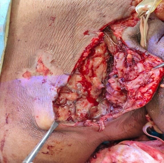
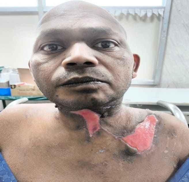
A case of necrotizing fasciitis
of neck:
A rare but life-threatening presentation of
necrotising fasciitis of the neck in a 46-year-old male, caused by Klebsiella
pneumoniae. The patient presented with neck swelling, dysphagia, blackish skin
discoloration, and foul-smelling discharge. Rapid deterioration led to septic
shock, multi-organ dysfunction, and cardiac arrest, requiring ventilatory
support and intensive care. Management included extensive surgical debridement,
tracheostomy, targeted intravenous antibiotics based on sensitivity, and supportive
care. Remarkable clinical improvement was seen with multidisciplinary
treatment, and the patient was eventually discharged in stable condition.
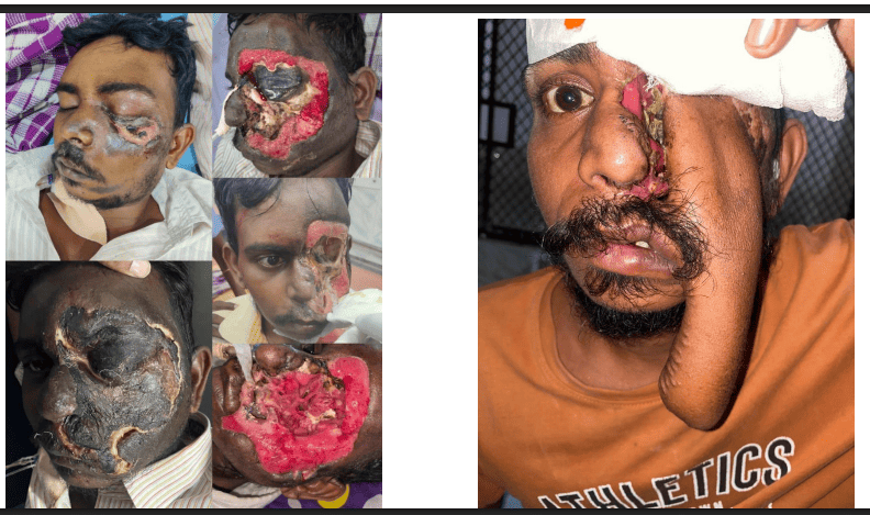
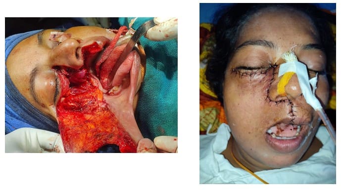
SUCCESS STORY 23:
Extensive rhinocerebral Mucor
mycosis with facial eschar:
A 31-year-old diabetic male presented with
facial swelling and black eschar following RTA. Imaging showed pansinusitis
with intracranial extension. Prompt endoscopic debridement with orbital
exenteration and liposomal Amphotericin B therapy and diabetic control to the
successful recovery of patient despite the high mortality associated with
cerebral involvement
SUCCESS STORY 24:
Post cricoid carcinoma in Young
female
An unusual presentation of a young female
presenting with progressive dysphagia, imaging revealing a large post cricoid
mass infiltrating the tracheal wall and thyroid gland. Biopsy confirmed as
moderately differentiated Squamous cell carcinoma. Case was presented in Tumour
board for definitive oncological management.
SUCCESS STORY 25:
Epithelioid angiosarcoma of nasal
cavity - In a young female
A 17-year-old female presented to us with
history of recurrent epistaxis, nasal obstruction and facial swelling.
Preoperative biopsy done in private medical college was positive for
angiofibroma. Imaging studies was throwing light on to differential diagnosis
as inverted papilloma, chronic fungal sinusitis with co-existing Antro choanal
polyp. Surgical excision of tumour was done by midfacial degloving approach and
histopathological was positive for a rare malignant vascular tumour -Epithelioid
angiosarcoma. Patient is under close oncological follow-up
SUCCESS STORY 26:
Odontogenic cyst- A maxillofacial
presentation
A 15-year-old female presented with painful
swelling in left maxillary region of 1-month duration. Imaging studies showed
well defined lesion around unerupted L canine. Patient underwent an endoscopic
Denker’s approach with medial maxillectomy- a minimally invasive procedure with
no facial incision or oroantral communication
SUCCESS STORY 27:
Osteosarcoma of left maxillary
sinus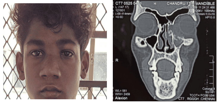
A 17-year-old male presented with rapidly
progressive swelling in left medial canthus and headache. Imaging showed lytic
lesion involving left medial maxillary sinus wall and posterior ethmoidal air
cells. Patient was managed with left middle metal antrostomy and mass excision via
endoscopic midfacial degloving approach. Patient is under close oncologic
follow-up up
SUCCESS STORY 28:
Spindle cell squamous carcinoma
of hard palate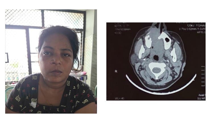
A 35-year-old female with history of hard
palate swelling and deep-seated pain. Imaging revealed infiltrating lesion
involving hard palate. Right total maxillectomy was done and HPE report
positive for Spindle cell squamous cell carcinoma and is given concurrent chemo
radiation.